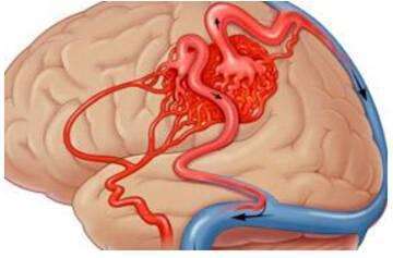
Venous malformation treatment methods
- EPA Announces First-Ever Regulation for “Forever Chemicals” in Drinking Water
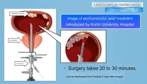
- Kochi University pioneers outpatient bladder cancer treatment using semiconductor lasers
- ASPEN 2024: Nutritional Therapy Strategies for Cancer and Critically Ill Patients
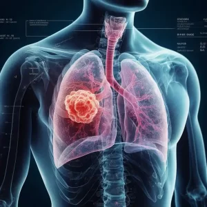
- Which lung cancer patients can benefit from neoadjuvant immunotherapy?
- Heme Iron Absorption: Why Meat Matters for Women’s Iron Needs
- “Miracle Weight-loss Drug” Semaglutide Is Not Always Effective
Venous malformation treatment methods
Venous malformation treatment methods. Venous malformations are not terrible. The choice of treatment is very important! The main treatment methods are interventional therapy, laser therapy, surgery and targeted drugs.
1. What is venous malformation?
Venous malformations, previously known as “cavernous hemangioma”, are composed of numerous sinusoids of endothelial cells. Sinusoids have different sizes and shapes, such as a sponge structure, hence the name. Venous malformations are one of the most common congenital vascular malformations, which can occur in any part of the body, with the craniofacial region and limbs being more common. Venous malformations cannot go away on their own, and can increase significantly with age. The main clinical symptoms are swelling, pain, bleeding, etc. If the lesion is located in an important functional area, it can affect the voice, swallowing and respiratory functions. In severe cases, it can die from bleeding and suffocation.
2. What examinations should be done for venous malformations?
Mainly ultrasound and magnetic resonance.
Color Doppler Ultrasound (CDFI) can objectively evaluate the flow rate, flow and other hemodynamic indicators of venous malformations. It is the first choice for screening. It can analyze the blood flow status of the diseased malformation vascular group, blood supply artery, and drainage vein And evaluation. In some cases that are difficult to locate, CDFI can also play a role in assisting positioning puncture.
Magnetic resonance can be used as the main examination method for diagnosing venous malformations, with extremely high sensitivity and specificity; it can accurately display the lesion area and the relationship with the surrounding soft tissues, and it is the best choice for diagnosis.
Compared with other auxiliary examinations, digital subtraction angiography (DSA) can visually and dynamically display the hemodynamic characteristics of the disease, and can also classify the disease. However, due to the invasiveness of digital subtraction angiography (DSA), it is generally completed at the same time as the interventional embolization treatment at this stage, and is rarely used as a preoperative evaluation and diagnosis method alone.
Foreign scholars divide it into 4 types according to the characteristics of venography:
- Type I: Isolated lesions are not accompanied by return veins;
- Type Ⅱ: the lesion drains to the normal return vein;
- Type Ⅲ: the lesion drains to the dilated and draining vein;
- Type IV: The lesion is an abnormally dilated vein;
Its classification is of great significance for treatment, especially interventional sclerotherapy.
3. What are the treatment methods for venous malformations?
The main treatment methods are interventional therapy, laser therapy, surgery and targeted drugs.
Mainly interventional therapy, mainly including sclerotherapy, embolization therapy, cryoablation and radiofrequency ablation. For type Ⅰ and Ⅱ lesions, sclerotherapy can obtain better clinical effects; for type Ⅲ and Ⅳ lesions, due to the faster flow of the return vein, sclerotherapy combined with embolization (coils, etc.) can improve the curative effect and reduce complications.
Among them, the advantages of sclerotherapy for venous malformations, such as simple treatment, quick onset, no local trauma, no severe necrosis, no scars, repetitive injection, and no obvious side effects, are being promoted continuously.
4. What is interventional sclerotherapy? What hardeners are there?
Interventional sclerotherapy is to puncture the venous tumor with a scalp needle or a puncture needle under the guidance of DSA. After the blood is drawn out, digital subtraction is performed to understand the scope of the tumor and the flow rate and shape of the drainage vein, and inject the sclerotherapy according to the imaging performance It can destroy the venous tumor body and its drainage vein.
Commonly used hardeners are:
- Absolute ethanol: after injection, it can cause protein denaturation, cause direct and extensive damage to endothelial cells, make it lose endocrine function, and promote the formation of abnormal blood vessel thrombosis. Anhydrous ethanol embolization therapy must be treated under general anesthesia to increase safety. Intraoperative vital signs such as invasive arterial pressure, blood oxygen saturation, and heart rate must be closely monitored.
- Polymer embolic agents: including NBCA and Onyx. Onyx is a non-viscous liquid embolic agent that is a mixture of vinylene alcohol polymer, dimethyl sulfoxide and tantalum powder. It can overcome the shortcomings of NBCA sticky tubes and is a more commonly used embolic agent. Polymer embolic agents have certain therapeutic significance, but because they do not have the effect of destroying vascular endothelial cells, the potential recurrence probability is relatively high.
- Mechanical embolization devices: such as occluders, coils, etc., although there is no direct chemical damage to the malformed veins, when the blood flow in the lesion is fast and the buffer is insufficient, the physical embolic devices can provide mechanical filling, thereby Slow down the flow rate, provide the possibility for further liquid embolism, and reduce the dose of liquid embolism. In the process of using absolute ethanol to treat malformed veins and return veins, full embolization with coils can reduce the flow rate of the disease, reduce the amount of absolute ethanol, improve treatment efficiency, and reduce the risk of system complications.
5. What are the side effects of interventional therapy? Will it recur?
The side effects of interventional therapy are mainly caused by sclerosing agents, especially absolute ethanol. Although absolute ethanol is the most effective and the only liquid embolic agent that can achieve the purpose of healing, its side effects are also the strongest. Complications that have been reported include nerve damage, skin ulcers, and fatal central cardiopulmonary failure.
The maximum single dose of absolute ethanol should not exceed 1 ml/kg to avoid hemolysis, acute kidney injury, heart and lung function accidents, etc. Due to the non-visualization of absolute ethanol, there are certain risks in the treatment process, and the addition of contrast agent will reduce its concentration and therapeutic effectiveness. Therefore, the absolute ethanol embolization treatment for high-flow venous malformations should be limited to those with rich experience and Professional physicians with a clear understanding of risk characteristics.
Due to the limited dose of the selected sclerosing drug, the location of the lesion and the volume of the tumor, multiple interventional sclerotherapy is usually required. The interval is about one and a half months. Generally, after the malformed vein and drainage vein are completely embolized, the chance of recurrence is very low.
(sourcechinanet, reference only)
Disclaimer of medicaltrend.org