X-ray CT MRI: What is the scope of examinations?
- EPA Announces First-Ever Regulation for “Forever Chemicals” in Drinking Water
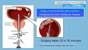
- Kochi University pioneers outpatient bladder cancer treatment using semiconductor lasers
- ASPEN 2024: Nutritional Therapy Strategies for Cancer and Critically Ill Patients
- Which lung cancer patients can benefit from neoadjuvant immunotherapy?
- Heme Iron Absorption: Why Meat Matters for Women’s Iron Needs
- “Miracle Weight-loss Drug” Semaglutide Is Not Always Effective
X-ray CT MRI: What is the scope of examinations?
- Red Yeast Rice Scare Grips Japan: Over 114 Hospitalized and 5 Deaths
- Long COVID Brain Fog: Blood-Brain Barrier Damage and Persistent Inflammation
- FDA has mandated a top-level black box warning for all marketed CAR-T therapies
- Can people with high blood pressure eat peanuts?
- What is the difference between dopamine and dobutamine?
- What is the difference between Atorvastatin and Rosuvastatin?
- How long can the patient live after heart stent surgery?
X-ray CT MRI: What is the scope of examinations? So what is the principle of X-ray inspection? Which diseases doctors will give priority to X-ray examinations?
Now everyone goes to the hospital without imaging examinations. X-ray, CT, and MRI are the most common imaging examinations.
Different examinations have different indications and contraindications, as well as different advantages, disadvantages and precautions.
There are many The public is not clear why these inspections are required. Let’s take a look at the differences between these inspections in detail!
01 X-ray photography
X-ray examination is commonly known as “filming”. So what is the principle of X-ray inspection? Which diseases doctors will give priority to X-ray examinations?
The principle of X-ray photography is that the density and thickness of human tissues are different. Therefore, when X-rays pass through different parts of the human body, the degree of X-rays absorbed is different, and the amount of X-rays reaching the screen or film will be different, forming light and dark or Contrast different images in black and white.
Ordinary X-ray photography is widely used in clinical practice because of its fast speed and low cost. However, X-ray is a two-dimensional image. There may be overlap of images in many parts, which affects the doctor’s observation and thus affects the diagnosis of the disease.
The display of subtle lesions is not clear enough, so for some subtle lesions, further examination by CT and MRI is required.
At present, the application of X-ray in tumor diagnosis and treatment mainly includes chest and abdomen plain radiographs, bone and joint photography, as well as gastrointestinal tract imaging and general breast X-ray photography.
The other inspection methods are less sensitive due to low sensitivity or no corresponding indications. . The current digital photography system DR can obtain high-quality images, and is widely used in orthopedic diseases (such as fractures), respiratory diseases (such as pneumonia, lung cancer), and abdominal diseases (such as intestinal obstruction, urinary calculi), etc. .
02 Computerized Tomography (CT)
CT actually uses the principle of X-ray, which can be regarded as an “advanced version” of X-ray. So what are the advantages and disadvantages of CT?
The principle of CT imaging is to use X-ray beams to continuously scan the tomography of a certain part of the human body. It has the characteristics of fast scanning time and clear images, and can be used for inspections of many diseases.
CT is generally divided into plain scan and contrast-enhanced scan. Plain scan refers to an ordinary scan without injection of contrast agent or contrast agent. It is mainly used for tissues with high contrast such as lungs and bones.
Most tumor lesions usually require enhanced scans to aid diagnosis. An enhanced scan is a method where iodide (such as iopamidol) is injected intravenously through a high-pressure syringe and then a CT scan is performed. Because the blood supply of each organ and lesion is different, the iodine concentration in each organ and lesion will be different, resulting in a density difference, which makes the lesion clearer.
CT is often used to examine the chest, abdomen, head, and bones and joints. Among them, chest CT has a clear structure, and its sensitivity and accuracy to chest lesions are better than conventional X-rays. Especially for the diagnosis of early lung cancer, chest CT is of decisive significance. It is of great significance for cancer patients to treat certain diseases such as interstitial pneumonia and pulmonary fibrosis.
CT is often used to examine the chest and abdomen of tumor patients. The structure of chest CT examination is clearer. The sensitivity and accuracy of detecting chest lesions are better than conventional X-ray chest radiographs, especially for the diagnosis of early lung cancer.
Chest CT is of decisive significance. It is of great significance for certain diseases such as interstitial pneumonia and pulmonary fibrosis caused by treatment for tumor patients, and the display of tumors in the mediastinum, lymph nodes and pleural diseases is also satisfactory.
Abdominal CT can clearly show the liver, gallbladder, spleen, pancreas, kidneys, adrenal glands and other organs of the solid organs. It can clearly show the accurate anatomy and the degree of lesions for tumors, infections and trauma, and has high value for the staging of lesions.
It is of high value for clinical The formulation of a treatment plan is of great significance especially for the surgical positioning of the operating department, and is of great value for the diagnosis and differential diagnosis of intra-abdominal masses. Head CT is the most routine and preferred examination method in traumatic craniocerebral emergencies.
It can clearly show brain contusion and laceration, acute intracerebral hematoma, epidural and subdural hematoma, craniofacial bone fracture, intracranial metal foreign body, arachnoid membrane Bleeding in the lower cavity and so on.
Although compared to X-ray tomography, CT is more expensive, but compared to X-ray, because it is a tomographic image, it can hierarchically display various differences in tissue, and the displayed tissue structure and the image of the lesion are compared with each other.
There is no overlap, which can significantly increase the detection rate of lesions, and at the same time perform three-dimensional reconstruction, which is convenient for doctors to observe and accurately locate. However, the diagnostic efficiency of CT examination for soft tissue tumors, especially the qualitative diagnosis, is not as good as MRI.
03 Magnetic Resonance Imaging (MRI)
Compared with the above two inspection methods, the principle of MRI is completely different, and it is also the most expensive of the three inspections. So what are its specific advantages and disadvantages?
Magnetic resonance imaging (MRI) distinguishes different tissues, including tumor tissues and normal tissues, based on the signal differences of protons in different compounds. MRI has been applied to the imaging diagnosis of diseases of various systems throughout the body.
The best results are the brain, spinal cord, cardiovascular, bone and joint, soft tissue and pelvic cavity. Its advantages are multi-sequence, multi-directional imaging, which can clearly observe the lesions from different angles, no ionizing radiation, good soft tissue imaging, and observation of lesions in the central nervous system, bladder, rectum, uterus, vagina, joints, muscles, etc. is better than CT .

For craniocerebral diseases, MRI is superior to CT because MRI has high soft tissue contrast and can accurately distinguish cerebral cortex (gray matter), medulla (white matter) and nerve nuclei, especially for diagnosis of medullary diseases, tumors, edema, etc.
The sensitivity is higher. In addition, MRI does not interfere with bone artifacts and is the preferred imaging method for diagnosing lesions in the pituitary gland, cranial nerves, brainstem, and cerebellum.
MRI application of contrast agent (enhanced MRI) can distinguish tumors and edema. However, for acute cerebral infarction, acute head trauma, etc., CT is generally checked first, and then MRI as needed.

The disadvantage of MRI is that it is unclear for moving organs such as the gastrointestinal tract, and the imaging effect is not good for the lungs and other parts lacking protons.
Magnetic resonance imaging is not as accurate and sensitive as CT in displaying calcification and bone lesions, and it is not suitable for people with metal objects in the body.
For example, people with pacemakers and contraceptive rings are not suitable for magnetic resonance examination because the examination takes a long time. , There is loud noise during operation, and patients with claustrophobic panic disorder are not suitable for examination.
X-ray CT MRI: What is the scope of examinations?
(source:internet, reference only)
Disclaimer of medicaltrend.org


