Nature: Australian scientists detect cancer by Smart microscope slides
- EPA Announces First-Ever Regulation for “Forever Chemicals” in Drinking Water
- Kochi University pioneers outpatient bladder cancer treatment using semiconductor lasers
- ASPEN 2024: Nutritional Therapy Strategies for Cancer and Critically Ill Patients
- Which lung cancer patients can benefit from neoadjuvant immunotherapy?
- Heme Iron Absorption: Why Meat Matters for Women’s Iron Needs
- “Miracle Weight-loss Drug” Semaglutide Is Not Always Effective
Nature: Australian scientists detect cancer by Smart microscope slides
- Red Yeast Rice Scare Grips Japan: Over 114 Hospitalized and 5 Deaths
- Long COVID Brain Fog: Blood-Brain Barrier Damage and Persistent Inflammation
- FDA has mandated a top-level black box warning for all marketed CAR-T therapies
- Can people with high blood pressure eat peanuts?
- What is the difference between dopamine and dobutamine?
- How long can the patient live after heart stent surgery?
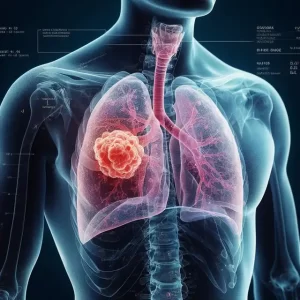
Nature: Australian scientists detect cancer by Smart microscope slides.
In a new study, Associate Professor Belinda S. Parker and Professor Brian Abbey of La Trobe University in Australia and their team found that by modifying the surface of traditional microscope slides at the nanometer scale, biological structures and cells will appear astonishing The color contrast can be used to detect diseases instantly.
The relevant research results were published in Nature on October 7, 2021. The title of the paper is “Colorimetric histology using plasmonically active microscope slides”.

Professor Abbey has developed this technology at La Trobe University in the past five years with co-inventor Dr. Eugeniu Balaur. Professor Abbey said, “Current tissue imaging methods often rely on staining or labeling cells in order to make them visible under a microscope.
Even with staining or marking, it is still challenging for pathologists to detect cancer cells, and some samples may be misdiagnosed, especially in the very early stages of the disease.
Recent breakthroughs in nanotechnology have allowed us to manipulate the interaction of light with biological tissues so that abnormal cells appear to have a different color from healthy cells.
Comparing the images on our slides with the traditional staining method, it’s like watching a color TV, and all you have seen before are black and white TV pictures. “
In this new research, Professor Abbey’s team and Associate Professor Parker’s team have tried to use this new technology called NanoMslide as an auxiliary method for diagnosing very early breast cancer.
Associate Professor Parker said that current technology can make it difficult to distinguish early forms of breast cancer from benign lesions, especially when there are not many abnormally shaped cells in complex tissues. NanoMslide makes this diagnosis easier.
Associate Professor Parker said, “When I observed the tissue under the microscope of NanoMslide for the first time, I was incredibly excited. For the first time I saw cancer cells suddenly appear in front of me. Their color is different from the surrounding tissues and it is very easy. Distinguish them from the surrounding cells.”
Associate Professor Parker believes that NanoMslide will complement the existing stains currently in use to achieve a more consistent cancer diagnosis. “Based on our preliminary research results on NanoMslide, we believe that this platform is really useful in the diagnosis of early breast cancer, and in other cancers, we really just want to single out some cancer cells in complex tissues or blood samples. “

Finite element method (FEM) optical transmission simulation, picture from Nature, 2021, doi:10.1038/s41586-021-03835-2.
Professor Abbey’s team developed their glass slide technology by leveraging the open access equipment and expertise provided by the Melbourne Nanomanufacturing Centre.
The team will work with the Melbourne Nanofabrication Center and the Australian National Fabrication Facility (ANFF) network to begin mass production of their slides to enter the market and solve a wide range of medical and non-medical imaging problems.
Professor John Dewar AO, Vice-Chancellor of La Trobe University, said that the invention of NanoMslide and its application in improving cancer diagnosis highlights the important role that universities like La Trobe University play in research innovation, and research innovation can improve The power of life.
Professor Dewar said, “As this outstanding invention transforms from an outstanding concept into a potentially life-saving solution, La Trobe University has proven that it can be achieved when outstanding research innovation is combined with a strong industry partner. What kind of results.”
Nature: Australian scientists detect cancer by Smart microscope slides
(source:internet, reference only)
Disclaimer of medicaltrend.org
Important Note: The information provided is for informational purposes only and should not be considered as medical advice.