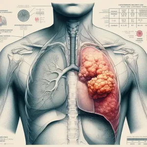
Israel: mRNA as Trojan horse can make cancer cells self-destruct
- Brief Intermittent Exercise Reduces Heart Disease and Death Risk
- Personalized Lung Tumor Chips Assess PD-1 Therapy Response
- Study Shows Prior Infection Offers Strong Immunity to Original COVID-19 Strain
- Chinese Food Products Dominate Korean Tables Amid Safety Concerns
- Early Detection of Hypopharyngeal Cancer Possible with Saliva Diagnosis
- EB Virus Could Be Infected by Kiss: A Hidden Threat Linked to Cancer
Israel: mRNA as Trojan horse can make cancer cells self-destruct
- AstraZeneca Admits for the First Time that its COVID Vaccine Has Blood Clot Side Effects
- Was COVID virus leaked from the Chinese WIV lab?
- HIV Cure Research: New Study Links Viral DNA Levels to Spontaneous Control
- FDA has mandated a top-level black box warning for all marketed CAR-T therapies
- Can people with high blood pressure eat peanuts?
- What is the difference between dopamine and dobutamine?
- How long can the patient live after heart stent surgery?
Israel: mRNA as Trojan horse can make cancer cells self-destruct.
Tel Aviv University in Israel recently issued a communiqué saying that researchers at the school successfully encoded the toxins produced by bacteria into mRNA (messenger ribonucleic acid) molecules and delivered them directly to cancer cells, and then induced these cells to produce toxins, thereby making cancer cells “suicide” .

The research results quickly caused a sensation on the Internet and media. The author consulted and sorted out the research papers published in Theranostics journal and the official website of Tel Aviv University, and let’s see what this so-called subversive discovery that allows cancer cells to self-destruct is what it is.
mmRNA-loaded liposomal nanoparticles

Figure: Schematic diagram of mmRNA-loaded liposome nanoparticles and their functions
The research team encoded the genetic information of a toxic protein produced by bacteria in the Pseudomonas family into Pseudomonas exotoxin mmRNA (similar to the method used to encode the genetic information of COVID-19’s “spike” protein into an mRNA molecule to create a vaccine). program).
The mmRNA molecules were then packaged in nanoliposome particles (LNPs) developed in the researchers’ lab to form mmRNA-loaded liposome nanoparticles (mmPE-LNPs), and the nanoliposomes were coated with antibodies to ensure the ability to produce the toxin.
Instructions are able to reach their target – cancer cells. Cancer cells swallow mmRNA-loaded liposome nanoparticles (mmPE-LNPs) through endocytosis, and release toxic mmRNA after intracellular dissolution of liposome nanoparticles, and mmRNA translates nanoliposome toxic proteins to lyse the cells to death .

Figure: In vitro therapeutic effect of mmPE-LNPs: reduction of cancer cell viability through apoptosis and protein translation inhibition
The researchers first investigated the antitumor ability of mmRNA-loaded liposome nanoparticles (mmPE-LNPs) in vitro.
After treatment with mmPE-LNPs in B16F10.9 melanoma tumor cell culture medium in vitro, it was observed that the proportion of apoptotic cells in B16F10.9 cells treated with mmPE-LNPs increased after 24 hours, and the proportion of apoptotic cells decreased after 48 hours.
Up to 90% (Figure D above). B16F10.9 melanoma cells were pre-incubated with mmPE-LNPs or mmFluc-LNPs for 2 hours, and then transfected with EGFP mmRNA by transfection reagent.
The results confirmed that EGFP expression was suppressed in mmPE-LNPs-pretreated cells compared with mmFluc-LNPs-pretreated cells, and this effect was dose-dependent (upper panel, B).

Figure: Effect of intratumoral injection of mmPE-LNPs on tumor growth in B16F10.9 mouse model
The study was followed by early animal testing. The study divided melanoma-bearing mice into three groups, and the mice in each group were injected with mmPE-LNPs (experimental group 0.15 mg/Kg), PBS (negative blank control), mmFluc-LNPs (mRNA control group) 4 times .
Results of the study showed that mice receiving mmPE-LNPs had smaller tumor volumes at all time points tested and significantly lower tumor volumes at the end of the experiment than controls (upper panel CD).

Figure: Effects of multiple intratumoral injections of mmPE-LNPs, PBS, and mmFluc-LNPs on B16F10.9 mice
Large-scale animal experiments continue to evaluate the effect of mmPE-LNPs on the survival of melanoma mice.
Mice inoculated with fluorescently labeled B16F10.9 tumor cells were still divided into three groups: mmPE-LNPs (experimental group 0.15 mg/Kg), PBS (negative blank control), mmFluc-LNPs (mRNA control group), each 2- Inject once every 3 days, and each mouse will be sacrificed when the tumor volume reaches 1500 mm3.
The experiment lasted 35 days from the inoculation of the tumor, with a total of 10 injection time points.
The study used the In Vivo Imaging System (IVIS) to track the intensity of the tumor every 2-3 days until the end of the experiment.
Results of the study showed that mice treated with mmPE-LNPs had reduced tumor volumes at all time points tested and significantly lower tumor volumes than controls at 20, 22, and 25 days after tumor inoculation (upper panel C).
In terms of survival rate OS, the experimental group and the control group began to separate at 20 days, and finally the survival rate of the mice in the experimental group was significantly better than that of the two control groups (Figure E above).
The in vivo imaging system (IVIS) tracked the treatment intensity of tumor infection and found that 44-60% of cancer cells had been infected by mmPE-LNPs (Figure F above) .
In addition, the study also evaluated the safety of mmPE-LNPs. The mmPE-LNPs did not induce major adverse effects of weight loss except by monitoring the body weight of the mice (top panel, B).
General toxicity was assessed by measurement of liver and spleen histology, liver enzyme levels, and pro-inflammatory cytokines in blood.
The results showed that compared with healthy mice, there were no significant changes in the tested tissues , and a single intratumoral dose of mmPELNPs (0.5 mg/Kg) had no effect on serum liver enzyme levels and IL-6 levels 24 hours after administration. Or the influence of TNF-α level .
Summary
The researchers innovatively encapsulated the reverse-compiled mRNA (mmRNA) of the Pseudomonas exotoxin A domain in nanoliposome particles (LNPs) to form mmRNA-loaded liposome nanoparticles (mmPE-LNPs), nano Liposome particles have a targeting effect on tumors because they are coated with antibodies.
After tumor cells swallow these nanoliposomes, they will release mmRNA and then translate and express toxic proteins to play an anti-tumor effect.
Through in vitro and animal experiments, it was found that mmPE-LNPs can infect 44%-60% of melanoma cells, mmPE-LNPs inhibited tumor growth and significantly increased the survival of melanoma mice.
In terms of safety, mmPE-LNPs had a slight effect on the body weight of mice, and the treatment did not cause any obvious systemic toxicity, nor did it cause abnormalities in immune factors.
Although mmPE-LNPs did have a very good curative effect in the first 20 days of treatment, it was found that the tumor began to recur after 22 days, and the tumor began to grow exponentially around 30 days (the red line in Figure C above), and the study has not yet found specific evidence of drug resistance. reason.
In conclusion, the innovative technology of liposomal nanoparticles (LNPs) encapsulating toxic protein mmRNA has indeed shown good efficacy and safety in anti-tumor, but the drug’s therapeutic window period and drug resistance mechanism have relatively large defects, which need to be further explored .
references:
Granot-Matok Y, Ezra A, Ramishetti S, Sharma P, Naidu GS, Benhar I, Peer D. Lipid nanoparticles-loaded with toxin mRNA represents a new strategy for the treatment of solid tumors. Theranostics. 2023 Jun 12;13(11 ):3497-3508. doi: 10.7150/thno.82228. PMID: 37441597; PMCID: PMC10334842.
Israel: mRNA as Trojan horse can make cancer cells self-destruct
(source:internet, reference only)
Disclaimer of medicaltrend.org
Important Note: The information provided is for informational purposes only and should not be considered as medical advice.