Immune Therapy-Induced Colitis: Deleting Fc Segment Emerges as a Solution
- Normal Liver Cells Found to Promote Cancer Metastasis to the Liver
- Nearly 80% Complete Remission: Breakthrough in ADC Anti-Tumor Treatment
- Vaccination Against Common Diseases May Prevent Dementia!
- New Alzheimer’s Disease (AD) Diagnosis and Staging Criteria
- Breakthrough in Alzheimer’s Disease: New Nasal Spray Halts Cognitive Decline by Targeting Toxic Protein
- Can the Tap Water at the Paris Olympics be Drunk Directly?
Immune Therapy-Induced Colitis: Deleting Fc Segment Emerges as a Solution
- Should China be held legally responsible for the US’s $18 trillion COVID losses?
- CT Radiation Exposure Linked to Blood Cancer in Children and Adolescents
- FDA has mandated a top-level black box warning for all marketed CAR-T therapies
- Can people with high blood pressure eat peanuts?
- What is the difference between dopamine and dobutamine?
- How long can the patient live after heart stent surgery?
Immune Therapy-Induced Colitis: Deleting Fc Segment Emerges as a Solution
Immune therapy has become widely used in anti-tumor treatment, but severe immune-related adverse events (irAE) have been a significant challenge hindering its effectiveness. Colitis is a common irAE, especially in the treatment with CTLA-4 inhibitors or their combination therapies.
However, the underlying mechanisms have not been thoroughly investigated in the laboratory, as mice rarely develop intestinal inflammation after immune therapy.
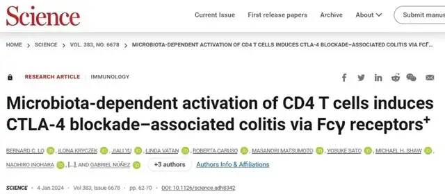
This week, a scientific team from the University of Michigan published a paper in the journal “Science,” revealing that CTLA-4 inhibitor-induced colitis is highly dependent on the intestinal microbiota. When researchers colonized the gut microbiota of wild-caught mice in laboratory mice, they exhibited immune characteristics closer to humans and showed significant colitis after immune therapy.
The study found that intestinal inflammation resulted from the dual action of over-activated CD4+ T cells producing IFN-γ and the depletion of regulatory T cells (Treg) induced by antibody Fc segment. Nanobodies lacking the Fc segment could prevent colitis without compromising anti-tumor effects.

As laboratory mice generally exhibit resistance to immune therapy-induced colitis, additional interventions, such as using dextran sulfate sodium (DSS) or introducing genetically susceptible mice to induce intestinal inflammation, were necessary to study the related issues.
Previous research indicated the inseparable relationship between the efficacy of immune therapy and the composition of the gut microbiota. Researchers speculated that laboratory mice used in experiments are usually bred in specific pathogen-free (SPF) environments, possibly leading to differences in their immune characteristics.
Researchers obtained reference gut bacteria (WildR) from wild-caught mice and attempted to introduce them into germ-free mice (GF) to establish an animal model of immune therapy-induced colitis. The results showed that WildR mice exhibited significant intestinal inflammation after receiving CTLA-4 and PD-1 inhibitor treatment, while mice colonized with SPF gut bacteria did not show colitis.
Further experiments revealed that the combination of CTLA-4 and PD-1 inhibitors, as well as the sole use of CTLA-4 inhibitors, could induce colitis in WildR mice, while the sole use of PD-1 inhibitors did not induce colitis, consistent with human studies.
Researchers analyzed immune cells in the cecum after CTLA-4 treatment and found an accumulation of CD4+ helper T cells (TH) expressing IFN-γ and IL-17, suggesting a significant TH1 response in immune therapy-related colitis. Simultaneously, researchers observed a decrease in Foxp3+ regulatory T cells (Treg) in the intestine, especially the selective depletion of RORγt+ peripheral induced Treg (pTreg) subpopulation. Previous studies found that RORγt+ pTreg had significant immune inhibitory features, such as overexpression of CTLA-4, IL-10, and bacteria-responsive TCR.
These results indicate that CTLA-4 inhibitors lead to TH1 response and depletion of RORγt+ pTreg in the intestine.
In clinical practice, immune therapy-induced colitis is often associated with tissue-resident cytotoxic CD8+ T cells. However, depleting CD8+ T cells did not affect colitis, while treating mice with IFN-γ neutralizing antibodies significantly alleviated intestinal inflammation during immune therapy.
It appears that intestinal inflammation induced by CTLA-4 inhibitors is primarily driven by CD4+ T cells and IFN-γ.
Due to the belief that Fc-mediated Treg depletion by anti-CTLA-4 antibodies is one of the mechanisms inducing tumor rejection, researchers speculated that this phenomenon might also be involved in the development of immune therapy-related colitis.
In mice lacking Fc receptors, CTLA-4 inhibitors did not cause intestinal inflammation, and no CD4+ T cells producing relevant cytokines were observed. Depletion of RORγt+ pTreg significantly improved the condition. This suggests that the Fc domain is essential for CTLA-4 inhibitor-induced colitis.
In turn, could the removal of the Fc segment solve this side effect?
Researchers chose an Fc-less CTLA-4 monoclonal antibody, H11, combined with a half-life extension agent (HLE). In mouse models of MC38 and CT26 colorectal cancer, H11-HLE demonstrated stable anti-tumor effects without inducing colitis.
Researchers speculated that the immunogenicity of free-living mouse gut bacteria is higher, and the immune tolerance threshold required for optimal anti-tumor immune response might be lower, making them more prone to intestinal inflammation under immune therapy.
In any case, identifying the key to CTLA-4 inhibitor-induced colitis opens up potential applications for further exploration.
Immune Therapy-Induced Colitis: Deleting Fc Segment Emerges as a Solution
References:
[1]https://www.science.org/doi/10.1126/science.adh8342
(source:internet, reference only)
Disclaimer of medicaltrend.org
Important Note: The information provided is for informational purposes only and should not be considered as medical advice.


