Research progress of transparent septum and related diseases
- Normal Liver Cells Found to Promote Cancer Metastasis to the Liver
- Nearly 80% Complete Remission: Breakthrough in ADC Anti-Tumor Treatment
- Vaccination Against Common Diseases May Prevent Dementia!
- New Alzheimer’s Disease (AD) Diagnosis and Staging Criteria
- Breakthrough in Alzheimer’s Disease: New Nasal Spray Halts Cognitive Decline by Targeting Toxic Protein
- Can the Tap Water at the Paris Olympics be Drunk Directly?
Research progress of transparent septum and related diseases
Research progress of transparent septum and related diseases. The transparent septum is a vertical membranous structure located in front of the midline of the brain. It has no nerve function. Due to the low incidence of lesions in this area, the existence of this structure and its related lesions rarely cause neurologists to pay enough attention.
The function of the transparent septum is not yet clear, but it may play an important role in the limbic system, and the relationship between its pathology and related mental disorders is also worthy of further study.
Based on the anatomical and developmental characteristics of the transparent septum, this article reviews some clinical features and treatment methods of related common diseases, and discusses the relationship between it and mental disorders.
1. Transparent septum anatomy and its developmental characteristics
1.1 Anatomical characteristics of transparent septum
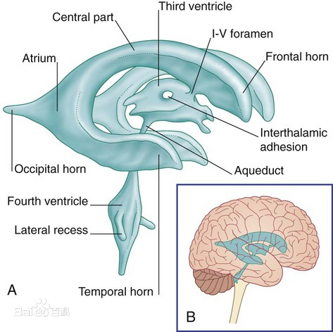
The transparent septum is a thin film composed of white matter fibers and a few neuronal tissues on the midline of the brain. It is located between the anterior horns of the two lateral ventricles and forms its inner wall. It extends in parallel to the interventricular foramen and separates the two lateral ventricles. open. The transparent septum is approximately triangular on the sagittal plane. The front and lower parts are the knees and mouth of the corpus callosum, the posterior and lower parts are the dome column, and the upper part is connected with the lower surface of the corpus callosum. The normal thickness of the transparent septum is 1~3mm. Its blood supply originates from the branch of the medial striatal artery of the anterior cerebral artery. Three septal veins run along the surface, and then cross the fornix column to enter the internal cerebral vein.
1.2 Developmental characteristics of transparent septum
The development of the transparent septum is closely related to the development of the corpus callosum. The corpus callosum is the connection structure of the white matter fibers between the two cerebral hemispheres. During the formation of the corpus callosum, when the corpus callosum extends to the cephalic side, the area between the corpus callosum and the fornix joint is pulled. Thinning to form a transparent compartment, the front joint plate gradually forms two transparent compartment lobules due to the “cavity effect”, and the cavity in it is a transparent compartment cavity. With the rapid development of the cerebral hemisphere and corpus callosum towards the head and tail, the transparent septal lobules on both sides gradually become thinner, and the transparent compartment cavity also becomes longer and wider, which lasts until the 24th week of pregnancy.
The vertical dome column divides the transparent compartment into the transparent compartment and the Verga cavity that communicate with each other back and forth. At 6 months of pregnancy, the two cavities were closed from back to front. At birth, 97% of the Verga cavity was closed and disappeared, and only the front transparent compartment was present. 3 to 6 months after delivery, 85% of the transparent compartment disappeared due to the fusion of the two diaphragms and became a transparent compartment, but the transparent compartment still exists in a few people in adulthood.
2. Hyaline septum dysplasia and hyaline septum cyst
The diseases related to the abnormal development of the transparent septum can be roughly divided into two categories: ① the absence of the transparent septum and its associated deformities. ② Transparent compartment cavity and transparent septal cyst.
2.1 Transparent septum and its associated deformities
The incidence of hyaline septum is 2~3/100,000. It is rare if it is simply absent. It is often accompanied by abnormal development of the forebrain and midline structure, such as dysplasia of the corpus callosum, dysplasia of the optic septum, and cleft brain malformation. Most of these lesions are Fatal or severe non-fatal central nervous system malformations. Therefore, it is very important to detect the absence of fetal transparent septum by prenatal ultrasound, which is of great significance for judging the abnormality of the fetal intracranial structure and timely termination of pregnancy.
2.2 Transparent compartment cavity and transparent septal cyst
The transparent compartment cavity mainly refers to the transparent compartment and Verga cavity that are still not closed after 3 to 6 months of birth. The Verga cavity often closes before the transparent compartment, so it is rare to exist alone. The transparent compartment often appears alone or occasionally combined with the Verga cavity.
Its existence can be without any symptoms for life, but some abnormally enlarged transparent compartments may cause some mental symptoms. SARWAR believes that when the two side walls of the transparent compartment are arcuate and the distance reaches 10mm, it is called a transparent septal cyst. The inner surface of the transparent compartment has no ependymal cells and is not connected to the cerebral ventricle.
The fluid in the cavity is formed by filtration through the diaphragm on both sides, and reabsorbed by the diaphragm vein and capillaries, and does not participate in the circulation of cerebrospinal fluid.
The formation mechanism of cystic fluid in hyaline septal cysts is still unclear. There are several hypotheses as follows: ① Congenital cysts may have the ability to secrete fluid from birth because of the variation of the cyst wall. ②Acquired cysts may obtain secretion ability through the differentiation of ependymal precursor cells or the migration of ependymal cells in the ventricle. Asymptomatic hyaline septal cysts generally do not require treatment. For hyaline septal cysts with clinical symptoms, the surgical indications and the best surgical method are still clinically controversial.
In general, the lesions that cause hydrocephalus or nerve compression with obvious symptoms and clear imaging diagnosis of lucid septal cysts should be treated with surgery, but there are also reports that hyaline septal cysts can be relieved by conservative treatment. Symptoms related to hyaline septum cysts (such as headaches, mental disorders, etc.) are non-specific.
Therefore, it is very important to correctly determine whether the symptoms are caused by asymptomatic hyaline septum cysts combined with other intracranial diseases or the clinical manifestations of hyaline septum cysts themselves. . The surgical treatment of hyaline septal cysts currently mainly include direct craniotomy and intracerebroventricular resection or fistula of hyaline septum cyst. Which method can better alleviate the clinical symptoms caused by hyaline septal cysts, there is no controlled study.
3. Transparent septal tumor
Tumors that originate in the transparent septum are very rare. Tumors in the transparent septum are more likely to be caused by the invasion of adjacent structures (usually the corpus callosum). The neoplastic lesions of the transparent septum are usually divided into primary tumors (which may originate from the transparent septum and the ependyma on its outer surface) and secondary tumors (from the surrounding structures of the transparent septum or metastases).
Central neurocytoma is the most common primary tumor. It has a high incidence in young people. It usually occurs in the transparent compartment near the interventricular foramen. It grows mainly from one side to the opposite side, and it often manifests as a broad base and lateral A mass connected to the hyaline septum of the ventricle with varying degrees of hydrocephalus generally has a good prognosis. In the 2016 WHO classification of the central nervous system, it was classified as neuron and mixed neuron-glial tumors, but the central nervous system Whether the cell tumor originated from the hyaline septum tissue or the neuronal tissue in the lateral ventricle is still unclear. In addition, there are subependymal giant cell astrocytoma and subependymoma in primary tumors. Among the secondary tumors, glioblastoma derived from the corpus callosum and primary central nervous system lymphoma are common; in addition, rare cases such as germ cell tumors and metastatic melanoma have also been reported.
Tumors that occur in the transparent septum are difficult to detect early. When the patient has obvious symptoms of increased intracranial pressure such as headache and vomiting, the tumor volume is often already very large. Like most intracranial tumors, surgery can clarify the nature of the disease and relieve it. Symptoms of increased intracranial pressure can improve the prognosis of patients. Sometimes patients may be accompanied by emotional or mental disorders in the early stage, but whether these symptoms are related to the pathology of the transparent septum structure itself is not yet clear.
4. Transparent septal lesions and mental disorders
The clinical symptoms caused by hyaline septal lesions, such as headache, decreased vision, etc., can be explained by imaging to suggest hydrocephalus or visual path compression, but the relationship between it and mental disorders has always been clinically significant. dispute. As tumor lesions in the lucid septum usually have damage to the surrounding structures (including the corpus callosum and vault, etc.), the lucid septum is generally associated with the developmental abnormalities of other structures in the forebrain or midline. At present, the gap between lucid septal lesions and mental disorders Most of the research focuses on the transparent compartment cavity (including transparent septal cyst, collectively referred to as the transparent compartment cavity below) population.
Transparent compartments can appear in depression, affective psychosis, schizophrenia and other mental disorders. SARWAR’s research on the embryology of the transparent septum and its fibrous connection believes that the septum is an important relay station between the hippocampus and the hypothalamus, and is a part of the limbic system. ALDRENTURNER believes that the transparent septum can be regarded as a relevant center that transmits visceral information to the hippocampus, amygdala, habenula, and brainstem network through the hypothalamic autonomic nervous system, and participates in consciousness, sleep processes and emotional responses.
Taking into account the functions of the limbic system in affective behavior, intentional instincts, and learning and memory, the appearance of the transparent compartment seems to partially explain some of the patients’ symptoms of mental disorders. However, the pathogenesis of schizophrenia has not yet been elucidated, and there is still considerable controversy as to whether it is a functional disease or an organic disease. Some studies believe that the incidence of transparent compartments in patients with schizophrenia is higher than that of the normal population, especially the large transparent compartments (the distance between the two side walls is greater than 6mm) may be the developmental abnormality of the limbic system of schizophrenia. A performance.
A large study analyzed a total of 1432 patients with schizophrenia and 333 healthy people. It is believed that the transparent compartment is related to abnormalities in the interpersonal relationship and emotional characteristics of mental patients. It also supports that the transparent compartment may be its brain. One of the structural characteristics of structural hypoplasia. However, some studies do not support the above conclusions. It is believed that there is no difference between the occurrence of larger transparent compartments between schizophrenia patients and healthy controls, and it is believed that the transparent compartments will not cause mental disorders.
In 2018, the author’s department performed neuroendoscopic fenestration on a female hyaline septum cyst with a significant depression (but no diagnosis of depression-related mental illness). After the operation, the depression of the patient was significantly relieved. In the following year During the follow-up, the patient’s family also reflected that the patient’s personality gradually became cheerful and optimistic after the operation. It is reported in the literature that a case of male hyaline septum cyst with memory loss and cognitive impairment has been significantly relieved after neuroendoscopic fenestration.
Can these cases suggest that the psychiatric symptoms caused by a hyaline septum cyst may simply be due to the pressure of the hyaline septum cyst itself on its surrounding structures (such as the corpus callosum, fornix, hypothalamus, etc.), because surgery cannot change the developmental abnormalities that exist in other brain structures. The dysplasia of the transparent septum and its related diseases are still relatively blank areas in clinical research.
Whether some tumors in the transparent compartment (such as central neurocytoma) originate from the transparent septum tissue; whether the abnormal transparent septum can be a cause of mental disorders; whether the transparent compartment cavity is related to the onset of schizophrenia; and whether the transparent compartment is related to the onset of schizophrenia; The symptoms of mental disorders related to the compartment cavity are due to the pressure of the transparent compartment cavity on the surrounding tissue structure, or because the existence of the transparent compartment cavity is only a manifestation of the developmental abnormalities of other structures of the brain; the solution of these problems requires the anatomy of the transparent compartment Further exploration of functions and larger-scale clinical controlled studies.
(source:internet, reference only)
Disclaimer of medicaltrend.org


