Application of tumor organoids in the evaluation of the efficacy of anti-angiogenic drugs
- Normal Liver Cells Found to Promote Cancer Metastasis to the Liver
- Nearly 80% Complete Remission: Breakthrough in ADC Anti-Tumor Treatment
- Vaccination Against Common Diseases May Prevent Dementia!
- New Alzheimer’s Disease (AD) Diagnosis and Staging Criteria
- Breakthrough in Alzheimer’s Disease: New Nasal Spray Halts Cognitive Decline by Targeting Toxic Protein
- Can the Tap Water at the Paris Olympics be Drunk Directly?
Application of tumor organoids in the evaluation of the efficacy of anti-angiogenic drugs
Application of tumor organoids in the evaluation of the efficacy of anti-angiogenic drugs. Tumor angiogenesis refers to the process of tumor cell-induced microvessel growth and the establishment of blood circulation in the tumor microenvironment.
It plays an important role in tumor growth, invasion and metastasis. It is also a hot spot for tumor research and new anti-cancer drug development [1 ].
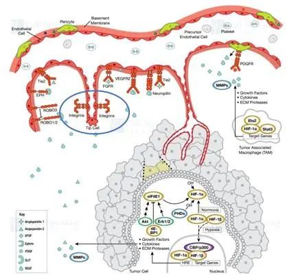
Figure 1: Signal pathway for angiogenesis (quoted from CST)
Common methods of in vitro evaluation of blood vessels
(1) 2D vascular endothelial cell detection: human umbilical vein endothelial cells (HUVEC cells) are planted in a cell culture dish to form a grid-like cavity structure to evaluate the effect of stimulating drugs on the proliferation of vascular endothelial cells or vascular morphology.
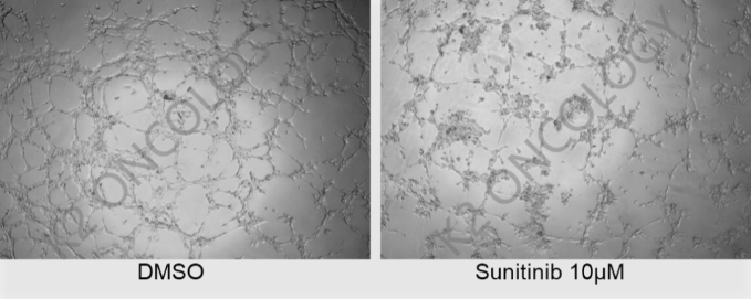
Figure 2: 2D vascular endothelial cell proliferation and blood vessel morphology detection
(2) 3D viscose beads angiogenesis detection: In 3D conditions, vascular endothelial cells are adhered to the fiber ball, and continue to be cultured on the matrigel ball. By observing and measuring the level of endothelial cell sprouting and luminal formation, qualitatively And quantitatively evaluate the influence of factors or drugs on angiogenesis.
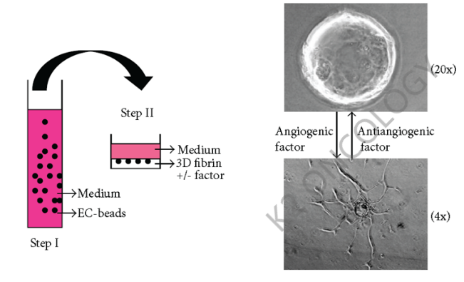
Figure 3: 3D viscose beads angiogenesis detection
Organoid and vascular endothelial cell co-culture model: 2D vascular endothelial cell detection and 3D viscose ball detection, neither provide the microenvironment in which tumor cells exist. The 3D co-culture model of organoids and vascular endothelial cells of Ketu Medical. The tumor organoids and vascular endothelial cells are co-cultured in vitro to form mixed culture spheres and then transferred to the surface of Matrigel to observe endothelial cell sprouting and luminal formation.
The advantages of this technical method are:
Provides a human-source three-dimensional co-culture model that is closer to the physiological state of tumor angiogenesis in vivo;
Established an organoid platform that can evaluate the efficacy of anti-tumor angiogenesis drugs/therapies.
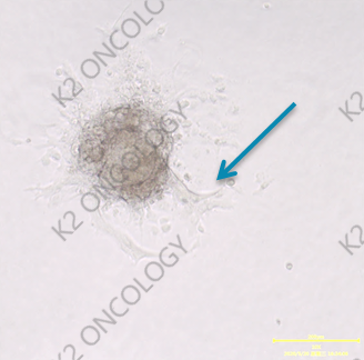
Figure 4: Co-culture model of tumor organoids and vascular endothelial cells
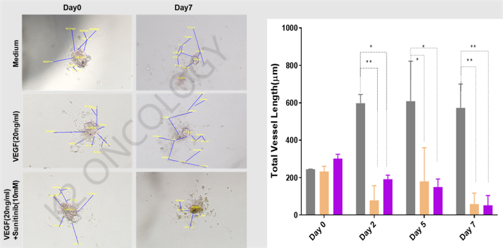
Figure 5: Co-culture of tumor organoids and vascular endothelial cells to evaluate drugs
The co-culture system of tumor organoids and vascular endothelial cells can effectively evaluate the ability of drugs to affect endothelial cell angiogenesis in multiple cancer types.
Through the 3D co-cultivation of tumor organoids, the three-dimensional co-culture system can truly reproduce the angiogenesis process of tumor organoids wrapped with vascular endothelial cells in vitro. The evaluation of drugs under this system is more authentic.
Ketu Medical’s 3D co-culture angiogenesis model bridges the gap between in vitro endothelial cells and animal models, and is a new and powerful tool for evaluating anti-angiogenesis drugs.
(source:internet, reference only)
Disclaimer of medicaltrend.org


