Unraveling HIV Persistence: Study Identifies Early Virus Reservoirs
- Normal Liver Cells Found to Promote Cancer Metastasis to the Liver
- Nearly 80% Complete Remission: Breakthrough in ADC Anti-Tumor Treatment
- Vaccination Against Common Diseases May Prevent Dementia!
- New Alzheimer’s Disease (AD) Diagnosis and Staging Criteria
- Breakthrough in Alzheimer’s Disease: New Nasal Spray Halts Cognitive Decline by Targeting Toxic Protein
- Can the Tap Water at the Paris Olympics be Drunk Directly?
Unraveling HIV Persistence: Study Identifies Early Virus Reservoirs
- Should China be held legally responsible for the US’s $18 trillion COVID losses?
- CT Radiation Exposure Linked to Blood Cancer in Children and Adolescents
- FDA has mandated a top-level black box warning for all marketed CAR-T therapies
- Can people with high blood pressure eat peanuts?
- What is the difference between dopamine and dobutamine?
- How long can the patient live after heart stent surgery?
Unraveling HIV Persistence: Study Identifies Early Virus Reservoirs
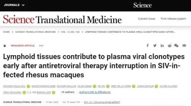
The original article titled “Contribution of Clones of Plasma Virus in Early SIV-Infected Macaques with Lymphoid Tissues to SIV Reservoirs after Antiretroviral Therapy Interruption” by Antonio Solis-Leal and colleagues was published on December 13, 2023, in “Science Translational Medicine,” a journal dedicated to translating scientific discoveries into practical medical applications.
Published by the American Association for the Advancement of Science (AAAS), the journal aims to promote interdisciplinary scientific research, particularly in the intersection between basic science and clinical applications.

Summary:
Scientists have made strides in understanding the rebound capability of certain HIV viruses during and after Antiretroviral Therapy (ART) interruption, a phenomenon known as the rebound-capable virus reservoir, posing a major challenge in HIV treatment.
To mitigate this impact, a study delved into the cells and tissues triggering virus rebound post ART interruption, focusing on the origin of the viral population.
By infecting macaques with barcoded SIVmac239M virus strains, the research analyzed virus clones in plasma and various tissues, revealing that secondary lymphoid tissues, such as mesenteric lymph nodes, could be a primary source of detectable viruses in the early stages.
Quantitative and qualitative analysis of virus DNA and RNA using techniques like single-cell RNA sequencing, RNAscope, and immunofluorescence provided insights into virus distribution and potential mechanisms in different tissues. These findings contribute to a better understanding of the reasons for HIV treatment failure and offer crucial clues for developing new therapies to prevent virus rebound.
Introduction:
The study highlights that despite prolonged Antiretroviral Therapy (ART), certain HIV viruses persist, capable of rapid resurgence after treatment interruption (ATI), leading to an increase in viral load. This rebound-capable virus reservoir poses a significant obstacle to achieving a cure for HIV. While current ART regimens effectively prevent new infections, they are powerless against dormant HIV viruses already present in the body.
Upon ART cessation, dormant viruses may become active again, causing viral spread and increased viral load. Uncontrolled virus activity can lead to immune suppression, AIDS, and a risk of transmission to others. Understanding the reasons, timing, and mechanisms of virus rebound after treatment interruption is crucial for developing new treatment strategies to minimize or eliminate these virus reservoirs.
The rebound-capable virus reservoir may include cells carrying HIV viruses capable of reactivation and causing effective infections. Besides lymphoid tissues, non-lymphoid tissues like the reproductive tract, fat, lungs, and brain, though having fewer latent infection cells, could also become sources of renewed HIV infection. Lymphoid tissues, especially those rich in CD4+ T lymphocytes, become significant reservoirs during suppressive ART.
Human lymphoid tissues are diverse, complex in structure and function, with approximately 800 lymph nodes in the body. It is speculated that during ATI, lymphoid tissues harboring replicable HIV viruses near CD4+ T cells may be a major source of HIV rebound. Analyzing specific locations of virus rebound in individuals with HIV theoretically has value, but due to sampling challenges, it is practically almost impossible.
Most current studies primarily analyze rebound viruses in peripheral blood, but blood-borne viruses may be a mixture from various tissues. Therefore, comprehensive research into all possible sources of HIV rebound viruses during early ATI is urgently needed.
To better understand the origins of early virus rebound after ART interruption, the researchers utilized a macaque model, allowing sampling of a wide range of tissues. In this study, a special simian immunodeficiency virus (SIV), named SIVmac239M, was used. This virus was genetically modified, incorporating a barcode consisting of 34 random nucleotides between the vpx and vpr genes.
This virus repository contained over 10,000 different virus barcodes, enabling tracking and analysis of virus clones at different time points in blood and tissues. These barcodes had similar replicative abilities, maintaining stability during extended replication. This method has been used for quantitative analysis and description of virus clones appearing in plasma after treatment interruption.
In this study, by deep sequencing early ATI samples, the researchers compared virus barcodes detected in peripheral blood, secondary lymphoid tissues, and non-lymphoid tissues during autopsy, identifying overlapping barcodes between plasma and tissues, suggesting these tissues might be key sources of virus reactivation and rebound after ATI.
Results:
The study assessed longitudinal plasma virus loads during SIV infection, ART, and ATI. The chronic plasma virus load of SIV-infected macaques was monitored for an extended period. In the second week post-infection, plasma virus levels peaked between 6.3 million and 27 million copies/mL. By the 12th week, when ART was initiated, plasma virus loads stabilized between 1,200 and 26,000 copies/mL.
In this study, chronic plasma virus loads in Chinese macaques were lower than those reported for Indian macaques infected with SIVmac239M, aligning with previous comparisons of SIVmac lineage virus responses between China and India. After 42 weeks of ART, some macaques showed slight virus fluctuations at specific time points (e.g., 20, 24, 28, 36, and 40 weeks), but most of the time, plasma virus loads remained below the detection limit of 81 copies/mL.
At specific stages of ART (28, 32, 36, and 40 weeks) and during the second ATI, more sensitive detection methods still showed virus loads below 22 copies/mL. After the first ATI (week 53), plasma virus RNA levels ranged from below 22 to 103 copies/mL. By week 57, plasma virus RNA levels rose to 347 to 20,000 copies/mL.
Following a brief 3-week treatment interruption, resumption of ART led to a rapid decrease in plasma virus load to below 81 copies/mL, maintained below 22 copies until euthanasia and autopsy (weeks 68 to 75), conducted within a week after the second ATI.
Unraveling HIV Persistence: Study Identifies Early Virus Reservoirs
References:
SCIENCE TRANSLATIONAL MEDICINE 13 Dec 2023 Vol 15, Issue 726 DOI: 10.1126/scitranslmed.adi9867
(source:internet, reference only)
Disclaimer of medicaltrend.org
Important Note: The information provided is for informational purposes only and should not be considered as medical advice.


