Heart Attack Spurs Cancer Growth: Heart’s Repair Vesicles Promote Tumor Growth
- Normal Liver Cells Found to Promote Cancer Metastasis to the Liver
- Nearly 80% Complete Remission: Breakthrough in ADC Anti-Tumor Treatment
- Vaccination Against Common Diseases May Prevent Dementia!
- New Alzheimer’s Disease (AD) Diagnosis and Staging Criteria
- Breakthrough in Alzheimer’s Disease: New Nasal Spray Halts Cognitive Decline by Targeting Toxic Protein
- Can the Tap Water at the Paris Olympics be Drunk Directly?
Heart Attack Spurs Cancer Growth: Heart’s Repair Vesicles Promote Tumor Growth
- Should China be held legally responsible for the US’s $18 trillion COVID losses?
- CT Radiation Exposure Linked to Blood Cancer in Children and Adolescents
- FDA has mandated a top-level black box warning for all marketed CAR-T therapies
- Can people with high blood pressure eat peanuts?
- What is the difference between dopamine and dobutamine?
- How long can the patient live after heart stent surgery?
Heart Attack Spurs Cancer Growth! Scientists Discover that Heart’s Repair Vesicles Released after Heart Attack Unexpectedly Promote Tumor Growth
Clinical data shows a correlation between myocardial infarction or heart failure and an increased risk of cancer, but the mechanisms behind this are unclear. Many believe this might be due to shared risk factors such as obesity, smoking, and type 2 diabetes.
The truth, however, is not so simple.
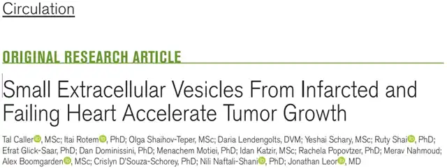
Recently, a paper published in the journal Circulation for the first time revealed this shocking fact: after a heart attack, the heart releases extracellular vesicles for cardiac repair, which unexpectedly promote tumor growth.

Jonathan Leor and colleagues from Tel Aviv University in Israel found that after a heart attack, cardiac mesenchymal stromal cells release a large number of extracellular vesicles to repair the heart. However, the biological molecules they carry also have tumor-promoting properties. When these extracellular vesicles circulate through the blood and reach malignant tumors or precancerous tissues, they promote the proliferation and migration of tumor cells.
A commonly used drug for cardiovascular disease can reduce the formation of these tumor-promoting extracellular vesicles, thereby inhibiting the accelerated growth of tumors after a heart attack.
Extracellular vesicles released by cells are enclosed in a double-layered membrane and contain various biomolecules such as RNA, cytokines, chemokines, growth factors, etc. These vesicles are an important means of transferring molecular biological information between cells and can also mediate communication between the heart and other organs. Therefore, extracellular vesicles may be a potential mechanism linking heart disease and cancer.
In this study, researchers first induced myocardial infarction in a mouse model of lung adenocarcinoma and found that in the early stage of left ventricular dysfunction (LVD) after the heart attack, the mouse heart produced a large number of extracellular vesicles, more than twice the amount in the control group (mice without myocardial infarction and with tumors).
The researchers decided to check the cargo sent out by the heart—surprisingly, they found that the extracellular vesicles produced by the heart after a heart attack actually had the characteristics of promoting tumor proliferation and migration!
Tracing back, the extracellular vesicles released by the heart after a heart attack mainly come from cardiac mesenchymal stromal cells (MSCs) that are activated during tissue damage and repair, hereinafter referred to as cMSC-sEV.
Identifying their cargo, compared to the extracellular vesicles released by the hearts of mice without myocardial infarction, cMSC-sEVs have a unique proteomic profile, rich in bone morphogenetic protein, osteopontin, IL-6, human galectin-3, TNF-α, VEGF, and other pro-inflammatory and pro-tumor cytokines, as well as miR-221, miR-214, and other pro-tumor-related microRNAs (a class of short non-coding RNA molecules that regulate gene expression).
Further validation through in vitro experiments showed that cMSC-sEVs promote tumor cell proliferation and migration in a dose-dependent manner and are tumor-specific. Treatment with cMSC-sEVs can accelerate the proliferation and migration of lung cancer and colon cancer cells, but has a smaller effect on melanoma and breast cancer cell lines.
Moreover, the in vitro results also showed that the cargo encapsulated by cMSC-sEVs not only directly affects tumors but also transforms macrophages into a pro-tumor phenotype, promoting macrophages to participate in the biological processes of inflammation, immune regulation, angiogenesis, and extracellular matrix remodeling, all of which are favorable conditions for tumor growth and spread.
In fact, the cargo transported by cMSC-sEVs was originally intended as a supplement to the heart after a myocardial infarction, inducing cell proliferation and tissue repair. However, when labeled with fluorescent dyes, it was observed that in addition to the heart, cMSC-sEVs also traveled to various parts of the mouse body, distributed in the liver, kidneys, spleen, lungs, bones, and tumor tissues, especially in the lungs congested by heart failure, where cMSC-sEVs gathered. This also means that while cMSC-sEVs warm the damaged heart, tumors also benefit.
As a result, the researchers thought of using the enzyme inhibitor GW4869 to inhibit the release of extracellular vesicles by reducing the synthesis of the essential material (ceramide) for extracellular vesicles. The results showed that injecting GW4869 into the peritoneal cavity of lung adenocarcinoma mice every 48 hours (the control group was injected with DMSO) after myocardial infarction effectively reduced the level of extracellular vesicles in the heart tissue and significantly inhibited tumor growth after myocardial infarction.
Consistent with the in vitro experimental results, GW4869 had little effect on mammary cancer mice after myocardial infarction, indicating that the pro-tumor effect of cMSC-sEVs is tumor-specific.
Moreover, researchers found a similar effect with spironolactone, a commonly used drug for cardiovascular diseases. The results showed that spironolactone reduced the level of cMSC-sEVs in disease mouse models by 28%, inhibiting tumor growth induced by myocardial infarction. It is worth noting that spironolactone itself does not have an anti-tumor effect, and when administered to tumor-bearing mice without myocardial infarction, the volume or weight of the tumors does not change.
Spironolactone, as a drug used long-term to treat cardiovascular diseases, has been well-validated for its safety and tolerability. This mouse experiment provides evidence that the use of spironolactone after a myocardial infarction may not only improve heart function but also bring additional anti-tumor benefits.
It’s unexpected that there’s such a connection between myocardial infarction and cancer. The supplements sent to the heart have become a stepping stone for tumors!
This study not only deepens our understanding of the connection between cardiovascular disease and cancer but also provides a new treatment strategy, possibly by regulating the production and function of cMSC-sEVs to reduce the risk of cancer in patients with cardiovascular disease.
Heart Attack Spurs Cancer Growth: Heart’s Repair Vesicles Promote Tumor Growth
references:
[1]Caller, T., Rotem, I., Shaihov-Teper, O., Lendengolts, D., Schary, Y., Shai, R., Glick-Saar, E., Dominissini, D., Motiei, M ., Katzir, I., Popovtzer, R., Nahmoud, M., Boomgarden, A., D’Souza-Schorey, C., Naftali-Shani, N., & Leor, J. (2024). Small Extracellular Vesicles From Infarcted and Failing Heart Accelerate Tumor Growth. Circulation, 10.1161/CIRCULATIONAHA.123.066911. Advance online publication. https://doi.org/10.1161/CIRCULATIONAHA.123.066911s://www.ahajournals.org/doi/10.1161/CIRCULATIONAHA. 123.066911
(source:internet, reference only)
Disclaimer of medicaltrend.org
Important Note: The information provided is for informational purposes only and should not be considered as medical advice.


