Fatty liver promotes cancer metastasis to the liver through extracellular vesicles
- Normal Liver Cells Found to Promote Cancer Metastasis to the Liver
- Nearly 80% Complete Remission: Breakthrough in ADC Anti-Tumor Treatment
- Vaccination Against Common Diseases May Prevent Dementia!
- New Alzheimer’s Disease (AD) Diagnosis and Staging Criteria
- Breakthrough in Alzheimer’s Disease: New Nasal Spray Halts Cognitive Decline by Targeting Toxic Protein
- Can the Tap Water at the Paris Olympics be Drunk Directly?
Fatty liver promotes cancer metastasis to the liver through extracellular vesicles
- Should China be held legally responsible for the US’s $18 trillion COVID losses?
- CT Radiation Exposure Linked to Blood Cancer in Children and Adolescents
- FDA has mandated a top-level black box warning for all marketed CAR-T therapies
- Can people with high blood pressure eat peanuts?
- What is the difference between dopamine and dobutamine?
- How long can the patient live after heart stent surgery?
Fatty liver promotes cancer metastasis to the liver through extracellular vesicles.
Colorectal cancer (CRC) is the third most common malignant tumor and the second leading cause of cancer-related death worldwide, with approximately 900,000 deaths from colorectal cancer each year worldwide.
The liver is the most common metastatic site of colorectal cancer because of the unique connection between the intestine and the liver through the portal vein.
Liver-specific metabolic and immune microenvironments also support liver metastasis in colorectal cancer. Ultimately, 70% of colorectal cancer patients develop liver metastases, which are the leading cause of death.
Obesity and nonalcoholic fatty liver disease (NAFLD) are important risk factors for colorectal cancer. More than 650 million adults worldwide are obese, and the increased prevalence of NAFLD has also been linked to the obesity epidemic.
Increasing epidemiological evidence shows that fatty liver increases the occurrence of colorectal cancer liver metastases and local recurrence after resection of colorectal cancer liver metastases, thereby worsening the prognosis.
Similarly, in an animal model of colorectal cancer liver metastases, fatty liver enhanced the growth of metastatic liver tumors by altering the hepatic inflammatory and immune microenvironment.
These findings suggest that the mechanisms of metastasis may vary due to the heterogeneity of fatty liver tumor microenvironment (TME) , which may explain the different responses of different patients to cancer therapy.
Therefore, the disease management of liver metastases may differ between fatty liver patients and non-fatty liver patients.
Understanding the molecular mechanisms underlying cancer metastasis in fatty liver patients is urgently needed for effective management and targeted therapy of these patients.
On May 11, 2023, researchers from Cedars-Sinai Medical Center published a research paper entitled: Extracellular vesicles in fatty liver promote a metastatic tumor microenvironment in the Cell Metabolism journal .
Here, we demonstrate that extracellular vesicles (EVs) produced by hepatocytes in fatty liver promote oncogenic YAP signaling and an immunosuppressive microenvironment through miRNAs , thereby promoting the progression of colorectal cancer liver metastases .
Extracellular vesicles (EVs) are nanoscale vesicles secreted by cells, containing a variety of proteins, lipids, and miRNAs, which play an important role in various physiological metabolic processes .
In nonalcoholic fatty liver disease (NAFLD) , the production of hepatocyte-derived extracellular vesicles (EVs) increases with altered miRNA content.
Previous studies have shown that primary tumor-derived EVs can translocate to the liver, promoting the formation of a pre-metastatic niche and a pro-metastatic inflammatory response.
However, the contribution of EVs produced in the liver, especially under NAFLD conditions, to the formation of the pre-metastatic and pro-metastatic liver microenvironments that predispose to CRC liver metastasis has not been thoroughly investigated.
In this latest study, the research team found that fatty liver produces extracellular vesicles (EVs) containing cancer-promoting miRNAs and creates a pre-metastatic liver microenvironment and pro-metastatic liver microenvironment that predisposes colorectal cancer liver metastases. Microenvironment.
The results showed that fatty liver up-regulates the expression of Rab27a, which in turn promotes the production of EVs by hepatocytes. In the liver, these EVs transfer miRNAs (miR25, miR92, miR103) that regulate YAP signaling from hepatocytes to cancer cells to enhance YAP activity by inhibiting LATS2.
In colorectal cancer liver metastases with fatty liver, YAP activity is elevated, induces M2 macrophage infiltration, promotes cancer cell growth and immunosuppressive microenvironment through production of CYR61.
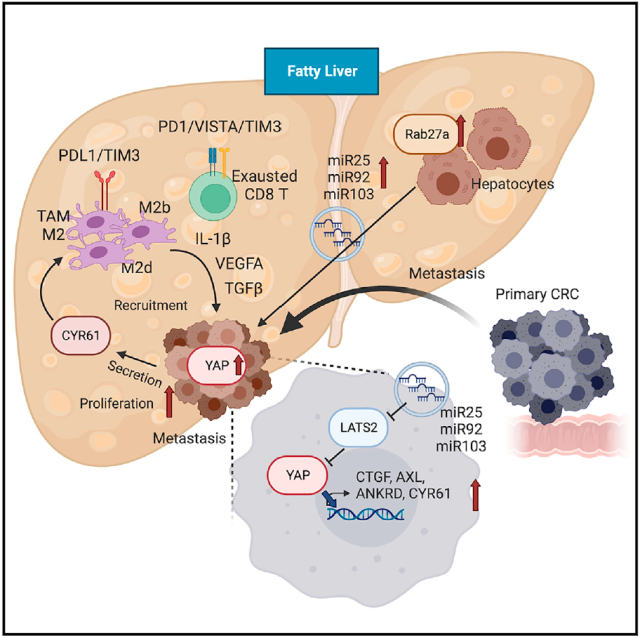
These results suggest that fatty liver-induced EV-miRNA, YAP signaling, and an immunosuppressive microenvironment promote CRC liver metastasis.
Therefore, nonalcoholic fatty liver disease (NAFLD) may generate a complex metastatic tumor microenvironment (TME) , which in turn promotes liver metastasis of colorectal cancer.
The increasing incidence of NAFLD worldwide in recent years may explain the differential response to cancer treatment in patients with colorectal cancer and liver metastases.
Paper link :
https://doi.org/10.1016/j.cmet.2023.04.013
Fatty liver promotes cancer metastasis to the liver through extracellular vesicles
(source:internet, reference only)
Disclaimer of medicaltrend.org
Important Note: The information provided is for informational purposes only and should not be considered as medical advice.


