Panoramic view of protein molecular pathology in patients with COVID-19
- Normal Liver Cells Found to Promote Cancer Metastasis to the Liver
- Nearly 80% Complete Remission: Breakthrough in ADC Anti-Tumor Treatment
- Vaccination Against Common Diseases May Prevent Dementia!
- New Alzheimer’s Disease (AD) Diagnosis and Staging Criteria
- Breakthrough in Alzheimer’s Disease: New Nasal Spray Halts Cognitive Decline by Targeting Toxic Protein
- Can the Tap Water at the Paris Olympics be Drunk Directly?
Panoramic view of protein molecular pathology in patients with COVID-19
Panoramic view of protein molecular pathology in patients with COVID-19. Cell report: Xihu University Guo Tiannan and others reveal the panorama of protein molecular pathology in patients with COVID-19 for the first time.
As of January 10, 2021, the global death toll caused by the new coronavirus infection has exceeded 1.93 million. We know that the virus can cause damage to multiple organs after invading the human body, but the molecular mechanism behind these damages is not clear, which limits the possible early intervention and treatment measures.
On January 8th, , Guo Tiannan’s research team and cooperative team (Hu Yu, Xia Jiahong, and Nie Xiu’s team from Union Hospital of Tongji Medical College, Huazhong University of Science and Technology) published the title “Multi-organ Proteomic Landscape” in Cell Online. of COVID-19 Autopsies,” the latest research paper reported a panorama of protein molecular pathology in multiple organ tissue samples from patients who died of new coronavirus pneumonia in early 2020. It is equivalent to they magnified the changes in the human body’s cell tissue after the COVID-19 that the doctor saw under the microscope by tens of thousands of times, reaching the protein molecular level, and “seeing” which molecular changes caused the disease and failure of human organs.
This is the first time in the world that, at the protein molecular level, a detailed and systematic analysis of the responses of multiple key organs after the new coronavirus infects the human body has been carried out, so as to formulate treatment plans and develop new novel coronaviruses for clinicians and researchers. The drugs and treatment methods provided clues and basis.
Link to the original paper:
https://www.cell.com/cell/fulltext/S0092-8674(21)00004-0
Or click “Read Original” at the end of the article to jump.
After being infected with the new coronavirus, 5336 protein molecules changed
A large number of clinical diagnosis and treatment and research have shown that the lungs and other organs of patients with new coronavirus disease have been damaged. However, most of the previous basic research related to COVID-19 used cell line models based on virus infection in the laboratory to speculate on the effects of the virus on various organs of the human body. There is a lack of pathological observation phenotypes for multiple organ damage in severely ill patients with COVID-19. The molecular level research behind it makes it difficult to deeply understand the death mechanism of the COVID-19 and to further carry out precise intervention treatment for patients.
The Guo Tiannan team of West Lake University and its collaborators collected tissue samples of seven organs (Figure 1) from the lung, spleen, liver, heart, kidney, thyroid, and testis of 19 patients who died from COVID-19. Through the pathological examination under the microscope, it can be found that the lungs of these patients have diffuse alveolar injury, pulmonary fibrosis, neutrophil infiltration and thrombosis and other pathological changes, spleen white pulp atrophy, liver fatty metaplasia and some cases Infarction occurred, myocardial edema and interstitial lymphocyte infiltration occurred in the heart, and acute tubular injury was found in the kidney.
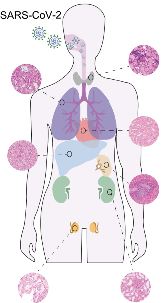
Figure 1. Multi-organ sample collection and microscopic pathological examination of patients with COVID-19
After that, research at the molecular level began. Based on high-pressure cycling technology (PCT) and TMT labeling combined with mass spectrometry data sampling and omics data analysis of shotgun proteomics technology, the research team identified 11394 human-source protein molecules and mapped out a panoramic view of multiple organ protein molecules in patients who died of COVID-19 critical illness (figure 2). Compared with control tissue samples of non-COVID-19 patients, 5336 proteins have changed (Figure 3).
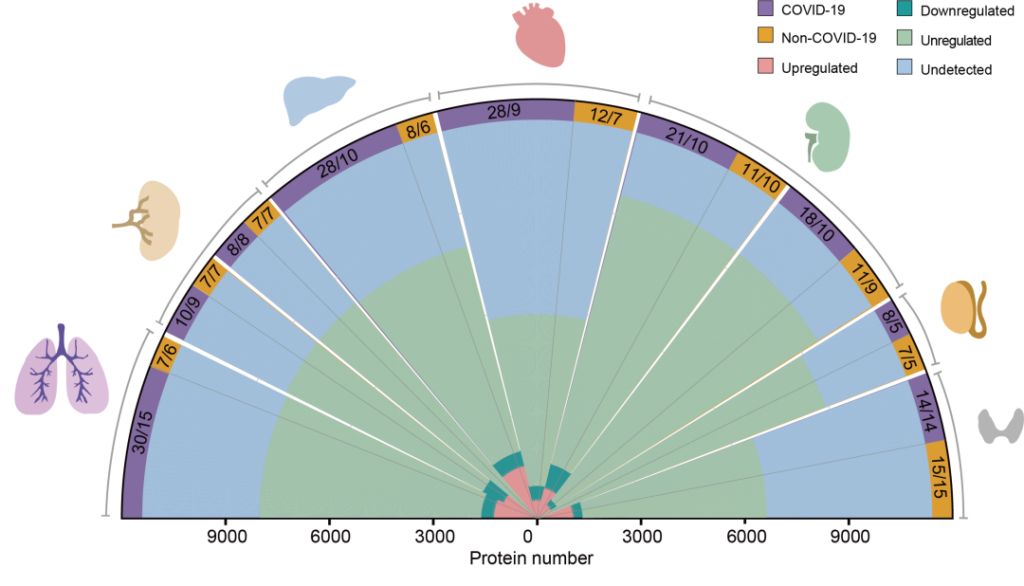 Figure 2. Schematic diagram of quantification of multi-organ proteome in patients with COVID-19
Among the seven organs and tissues of the human body, no significantly changed protein was identified in the red pulp of the spleen, while the number of changed proteins in the liver was the largest (N=1970), which means that the liver damage in patients with new coronavirus pneumonia may be more severe.
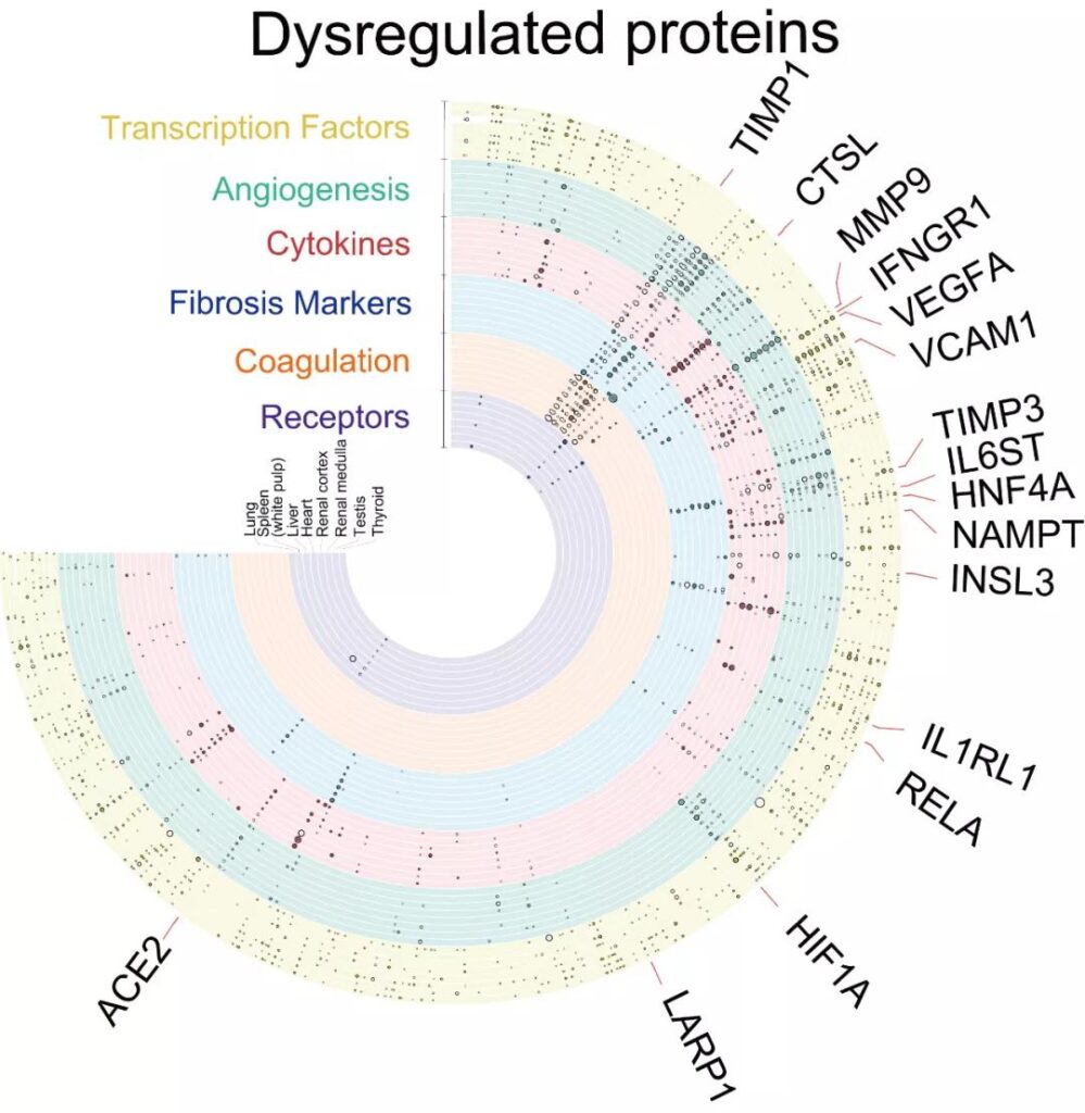
Regarding the “culprit” ACE2 protein (viral receptor angiotensin converting enzyme 2, a protein that regulates blood pressure in the human body), the “culprit” of the COVID-19 virus entering the human body, the research team found that its number is in the various organs of COVID-19 patients and non- COVID-19 patients. There is no significant difference.
Another protein, cathepsin L (CTSL), which helps the virus enter the cell, has been significantly increased in the lungs of patients with new coronavirus disease (Figure 4).
This suggests that the expression level of ACE2 has not changed in patients lethal to the new coronavirus. It is only a channel for the new coronavirus to enter the human body. CTSL may be a potential therapeutic target to block virus invasion.
Except the lungs, Liver and kidney also show signs of fibrosis
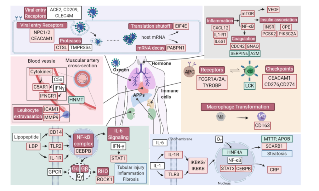
Figure 4. Schematic diagram of specific molecular changes related to the high inflammation state of multiple organs and tissue damage after the virus invades the human body.
The red/green boxes and black letters respectively indicate significantly up/down-regulated proteins, the red/green boxes and white letters indicate obviously activated/inhibited pathways, and the white boxes indicate unquantified or unregulated proteins.
The research team further carried out a systematic comparative study on the physiological functions, pathological morphology and proteomics of various organs (Figure 4), and found changes in multiple lung proteins, including those related to virus proliferation, involvement in the pathological process and degradation of lung fibrosis Viral restriction factor protein.
Proteomics also showed that the lungs and spleen showed up-regulation of immune checkpoint proteins and down-regulation of T cell enriched proteins as molecular features of adaptive immune response suppression, and the reduction of T, B and other lymphocytes in the spleen was also confirmed The molecular characteristics.
From the clinicopathological point of view, although only substantial fibrotic lesions occurred in the lungs, the proteomics results (Figures 3 and 4) showed that signs of tissue fibrosis were also observed in the liver, kidneys and other organs, suggesting that For critically ill patients with new coronavirus disease who have recovered, they need to prevent and take early intervention for the possible sequelae of “multiple organ fibrosis”.
The clinical collaborators in the research team reported for the first time in May 2020 that the testis of patients who died from the new coronavirus infection had seminiferous tubule damage, Leydig cell reduction, and mild lymphocytic inflammation.
But these only stay at the “macro” level. What molecular changes have caused these damages? Guo Tiannan’s laboratory found 10 proteins that have significantly changed in the testis tissue of patients with COVID-19.
Their functions are closely related to the inhibition of cholesterol synthesis, the reduction of sperm activity, and the reduction of Leydig cell-specific markers (Figure 5). Among them, Leydig cells are closely related to the synthesis and secretion of male androgen, suggesting that the fertility of male patients with COVID-19 may be affected.
Of course, these studies are based on tissue samples from patients who died from t COVID-19. Whether the same changes will occur in mild and severe patients, and whether such changes are reversible, still needs further research.
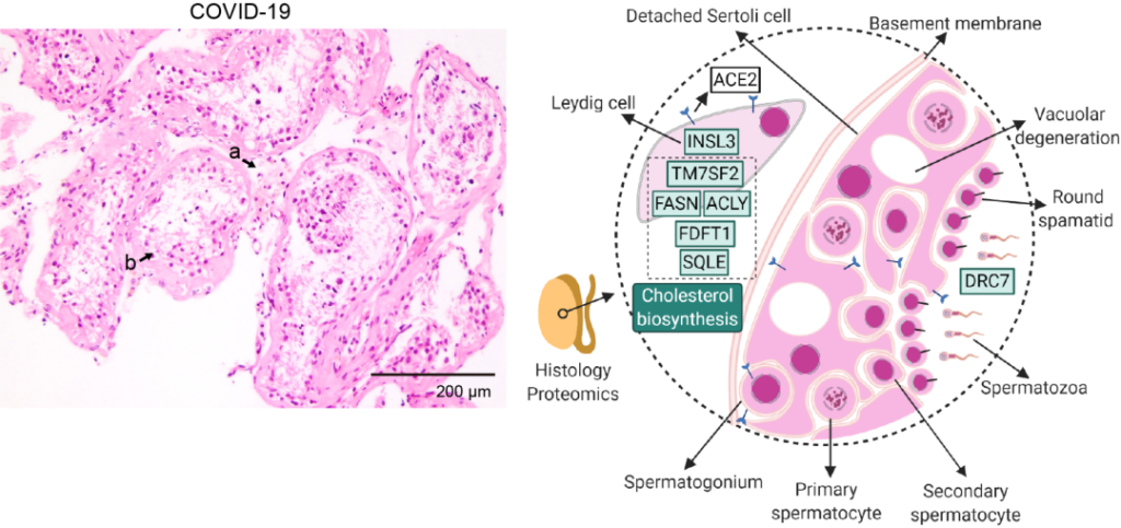
Figure 5. Schematic diagram of Leydig cells in the testicular interstitial space of patients with COVID-19 death, and the significantly down-regulated proteins and related pathways in the tissue.
The first batch of Chinese COVID-19 vaccines arrived in Uruguay
(sourceinternet, reference only)
Disclaimer of medicaltrend.org
Important Note: The information provided is for informational purposes only and should not be considered as medical advice.


