How to deal with cerebral aneurysm rupture?
- Normal Liver Cells Found to Promote Cancer Metastasis to the Liver
- Nearly 80% Complete Remission: Breakthrough in ADC Anti-Tumor Treatment
- Vaccination Against Common Diseases May Prevent Dementia!
- New Alzheimer’s Disease (AD) Diagnosis and Staging Criteria
- Breakthrough in Alzheimer’s Disease: New Nasal Spray Halts Cognitive Decline by Targeting Toxic Protein
- Can the Tap Water at the Paris Olympics be Drunk Directly?
How to deal with cerebral aneurysm rupture?
How to deal with cerebral aneurysm rupture? What happens when a cerebral aneurysm ruptures? What to do if it breaks?
What happens when a cerebral aneurysm ruptures? An aneurysm is a balloon-like bulge in the wall of an artery. As the aneurysm grows, it puts pressure on nearby structures and may eventually rupture. A ruptured aneurysm releases blood into the subarachnoid space around the brain. Subarachnoid hemorrhage (SAH) is a life-threatening stroke. The focus of treatment is to stop bleeding and repair aneurysms through clipping, coiling or bypass.
What is a ruptured aneurysm?
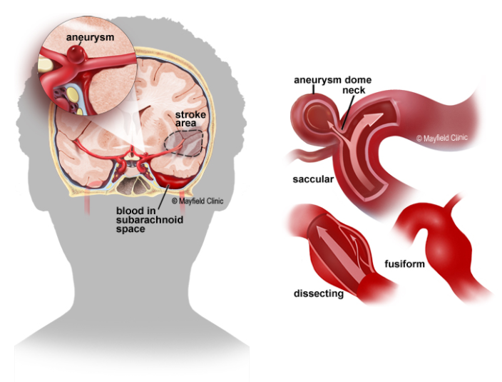
An aneurysm is a balloon-like bulge or weakening of an artery wall. (Similar to the balloon on the side of a garden hose.) As the bulge grows, it becomes thinner and weaker. It will become so thin that the blood pressure inside will cause it to leak or rupture. Aneurysms usually occur in larger blood vessels at the bifurcation of the artery. Types of aneurysms include (Figure 1):

Figure 1: A ruptured aneurysm releases blood into the subarachnoid space around the brain, causing a stroke (left). Different types of aneurysms (right). (What happens when a cerebral aneurysm ruptures?)
- Saccular-(most common, also called “berry”) aneurysms protrude from one side of the artery with a distinct neck at the bottom.
- Fusiform-the aneurysm swells in all directions without a distinct neck.
- Anatomy-A tear in the inner wall of an artery stratifies and collects blood; usually caused by trauma.
When an aneurysm ruptures, it releases blood into the space between the brain and skull. This space is filled with cerebrospinal fluid that soaks and buffers the brain. As the blood spreads and clots, it stimulates the lining of the brain and damages brain cells. At the same time, the areas of the brain that previously received oxygen-rich blood from the affected arteries are now deprived of blood, leading to strokes. Subarachnoid hemorrhage (SAH) is life-threatening, with a 40% risk of death.
The clotted blood and fluid collect in the hard skull and increase the pressure, which may squeeze the brain or cause the brain to shift and herniate. The obstruction of normal cerebrospinal fluid circulation can enlarge the ventricles (hydrocephalus), leading to confusion, lethargy, and loss of consciousness.
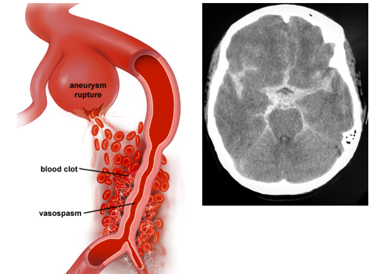
The complication that occurs 5 to 10 days after the aneurysm ruptures is vasospasm (Figure 2). Stimulating blood byproducts can cause spasms and narrowing of the arterial walls, reduce blood flow to this area of the brain, and cause secondary strokes.
 Figure 2: (Left) When red blood cells are broken down, toxins cause the walls of nearby arteries to spasm and narrow. The greater the SAH, the higher the risk of vasospasm. image 3. (Right) CT scan shows a ruptured aneurysm causing subarachnoid hemorrhage (white star). (What happens when a cerebral aneurysm ruptures?)
What are the symptoms of ruptured cerebral hemangioma?
Most aneurysms do not show symptoms until they rupture. Ruptures usually occur when a person is active rather than sleeping.
·Severe headache suddenly (usually described as “the worst headache in my life”)
·Nausea and vomiting
torticollis
·Sensitive to light (photophobia)
·Fuzzy or double vision
Loss of consciousness
·Epilepsy
What are the treatments for ruptured cerebral hemangioma?
Treatment may include life-saving measures, symptom relief, hemorrhage aneurysm repair, and prevention of complications. Within 10 to 14 days after the aneurysm ruptures, the patient will remain in the Neuroscience Intensive Care Unit (NSICU), where doctors and nurses can closely observe signs of rebleeding, vasospasm, hydrocephalus, and other potential complications.
Drugs
Pain medications will be used to relieve headaches, and anticonvulsants may be used to prevent or treat seizures.
Cerebral aneurysm rupture surgery
Determining the best treatment for a ruptured aneurysm involves many factors, such as the size and location of the aneurysm and the stability of the patient’s current condition.
- Aneurysm clipping: Cut an opening in the skull, called craniotomy, to locate the aneurysm. Place a small clip on the “neck” of the aneurysm to prevent normal blood flow from entering. The clip is made of titanium and remains permanently on the artery.
- Aneurysm embolization: It is done during angiography in the radiology department. The catheter is inserted into the artery in the groin and then through the blood vessel to reach the aneurysm in the brain. Through the catheter, the aneurysm is wrapped with platinum coils or glue to prevent blood from flowing into the aneurysm.
- Arterial embolism and bypass: If the aneurysm is large and inaccessible, or the artery is severely damaged, the surgeon may perform bypass surgery. Craniotomy opens the skull and clamps are placed to completely block (occlude) arteries and aneurysms. Then, by inserting a vascular graft, blood flows through the blocked artery. The graft is a small artery that is usually removed from your leg and attached above and below the blocked artery to allow blood to flow through the graft.
- Another method is to separate the donor artery from its normal position on the scalp and connect it to the blocked artery in the skull. This is called STA-MCA (superficial temporal artery to middle cerebral artery) bypass.
(sourceinternet, reference only)
Disclaimer of medicaltrend.org


