Why do women’s fertility decline after 35 years of age?
- Normal Liver Cells Found to Promote Cancer Metastasis to the Liver
- Nearly 80% Complete Remission: Breakthrough in ADC Anti-Tumor Treatment
- Vaccination Against Common Diseases May Prevent Dementia!
- New Alzheimer’s Disease (AD) Diagnosis and Staging Criteria
- Breakthrough in Alzheimer’s Disease: New Nasal Spray Halts Cognitive Decline by Targeting Toxic Protein
- Can the Tap Water at the Paris Olympics be Drunk Directly?
Why do women’s fertility decline after 35 years of age? New research found another important factor
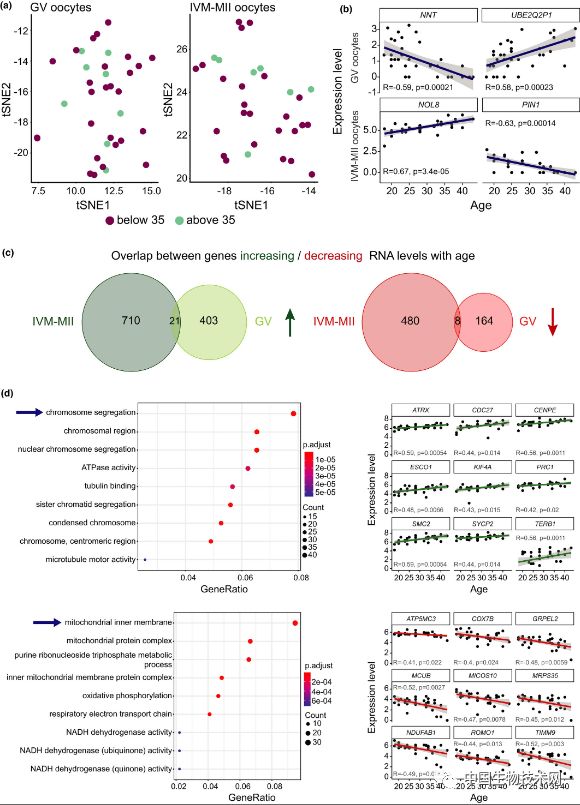
Why do women’s fertility decline after 35 years of age? In a new study published in “Aging Cell”, a research team led by the Center for Genome Regulation (CRG) of the Barcelona Institute of Science and Technology in Spain found that age may also affect women’s fertility by destroying the level of RNA molecules.
The RNA molecule changes the function of genes involved in key biological pathways in the final mature stage of human egg cells.
The oocyte (oocyte) is the oocyte that undergoes meiosis during oogenesis. Divided into primary oocytes, secondary oocytes and mature oocytes, they are the products of one, two and three meiosis respectively. At birth, the number of primary oocytes in the two ovaries of female babies reaches about 2 million, which are all the oocytes in their lives, and gradually decrease with age.
In this study, the researchers investigated the changes in the transcriptome associated with oocyte aging to understand which genes’ activity is affected by age. They used single-cell sequencing technology to analyze the transcriptome of 72 oocytes from 37 donors between the ages of 18 and 43.
The results showed that with age, the number of gene transcripts involved in chromosome separation and RNA processing gradually increased, while the number of transcripts related to mitochondrial metabolism gradually decreased.
The researchers found that the mature stage of oocytes is the main driver of transcriptome variation. During the growth process, oocytes have transcriptional activity. Once they reach the final stage of their growth and development, they become inactive, thus completing their nuclear maturation mission.
In immature egg cells, the transcriptome is less affected by age. The researchers believe that these findings indicate that age may affect the ability of oocytes to process gene products, which are critical to the last step of oocyte development.
Professor Bernhard Payer, the corresponding author of the study and director of the CRG, said: “Our research shows that the last step of oocyte maturation may itself be negatively affected by age, which is critical for fertility because it provides early embryos. Substances needed for normal development and survival. But what we don’t know is which of these changes are just the result of the aging process, and which of these may directly cause the quality of oocytes to decline with age.”
The researchers revealed some potential major regulatory genes through further analysis, which are also affected by age. The researchers said that future work will test whether these genes play a key role in oocyte aging.
The researchers also used the height and weight information of the donor to assess the impact of body mass index (BMI) on the transcriptome. The sample included oocytes from most women in the range of normal BMI (18.8-24.9) and overweight (BMI=25-30), as well as 1 underweight woman (BMI=17) and 1 obese woman (BMI=32). Like the age factor, the researchers regarded BMI as a continuous variable and looked at the correlation between BMI and antral follicle count (AFC) and BMI and gene expression. The results showed that, unlike age, BMI The abnormality mainly affects the transcriptome of immature oocytes. This finding suggests that the decline in fertility caused by age may have a different mechanism from the decline in fertility caused by abnormal BMI.
The researchers said that although more in-depth research is needed, this discovery will help develop new diagnostic tools in the future to better evaluate the quality of oocytes in reproductive medicine, as well as potential drug therapies to regulate The biological pathways that affect it, rejuvenate aging oocytes
(source:internet, reference only)
Disclaimer of medicaltrend.org


