New target for Alzheimer: Target Activation of PAC1R and Clear Toxic Protein
- Normal Liver Cells Found to Promote Cancer Metastasis to the Liver
- Nearly 80% Complete Remission: Breakthrough in ADC Anti-Tumor Treatment
- Vaccination Against Common Diseases May Prevent Dementia!
- New Alzheimer’s Disease (AD) Diagnosis and Staging Criteria
- Breakthrough in Alzheimer’s Disease: New Nasal Spray Halts Cognitive Decline by Targeting Toxic Protein
- Can the Tap Water at the Paris Olympics be Drunk Directly?
Science: New target for Alzheimer’s disease treatment, Target activation of PAC1R, and clear toxic protein
New target for Alzheimer: Target Activation of PAC1R and Clear Toxic Protein. The accumulation of pathological tau in synapses is related to the accumulation of ubiquitinated proteins.
Alzheimer’s disease (AD) is a common neurodegenerative disease. The accumulation of pathological tau protein in synapses has been identified as an early event of AD, and is closely related to the cognitive decline of AD patients. Synaptic dysfunction and loss of synaptic spine are also associated with cognitive decline in AD.
Under normal physiological conditions, tau protein is mainly concentrated in the cytoplasm and axons of neurons, and does not exist in a large amount in synapses. However, under disease conditions, due to the sorting error within neurons, the cross-neuronal For abnormalities in the transfer or clearance pathway, tau will accumulate in the postsynaptic compartment and presynaptic terminal.
Studies on mouse models of tau pathology have shown that hyperphosphorylation of tau can lead to mislocalization of tau on dendrites and dendritic spines.
Interestingly, there are studies claiming that the accumulation of pathological tau in synapses is related to the accumulation of ubiquitinated proteins, which suggests an abnormality in the synaptic ubiquitin proteasome system (UPS). The UPS mechanism in the synapse is essential for the normal function of the synapse, such as synaptic protein turnover, synaptic plasticity, and long-term memory formation.
In neurites, inhibiting protein degradation will lead to the wrong sorting of phosphorylated tau protein on dendrites and the loss of dendritic spines, while enhancing the protein degradation pathway will reduce the wrong selection of tau protein, which suggests that the protein degradation system is in the tau direction. The distribution of dendrites and dendritic spines plays an important role.
The degradation of tau and other polyubiquitinated substrates is accomplished by the 26S proteasome, which is a protease that can degrade proteins into small peptides; the decrease in proteasome activity and its effects have also been observed in AD patients. Changes in conformation. These data suggest that damage to proteasome activity may be a common mechanism in various neurodegenerative diseases.
In previous studies, related studies in the Natura Myeku laboratory at Columbia University Irvine Medical Center showed that the pure 26S proteasome in the brain of tau transgenic mice (rTg4510) has defects in degrading ubiquitinated proteins, while phosphodiester Enzyme inhibitors can enhance the activity of the proteasome by cyclically activated protein kinase (cAMP)/cAMP-dependent protein kinase A (PKA), the cAMP/PKA signaling pathway, to hydrolyze tau, thereby alleviating the pathology of tau protein and improving cognitive function .
However, activating or enhancing intracellular protein degradation through the proteasome or autophagy pathway may not be an ideal strategy for the treatment of AD. Therefore, in the early stages of AD, targeting tau at specific locations in neurons may be a safer treatment strategy to prevent tau accumulation, spread, and cognitive decline.
On May 26, 2021, Natura Myeku’s laboratory published a research paper titled “PAC1 receptor–mediated clearance of tau in postsynaptic compartments attenuates tau pathology in mouse brain” in the journal Science Translational Medicine.
The study found that PACAP1 receptors can reduce the amount of tau protein in the synapses of Alzheimer’s disease model mice, alleviate tau protein pathology, and improve the cognitive ability of mice.
In this article, first of all, the author found that as the disease progresses in tau transgenic mice (rTg4510) (Pre-, mid- and post-synaptic), the amount of pathological tau protein increases in neuron cell bodies, synapses, pre-synapses and postsynapses, especially in the cell bodies; and compared to pre-synaptic, post-synaptic A large number of different types of tau proteins have been accumulated, such as tau monomers, high molecular weight tau (65 kDa), and mutant tau (AT8) (Figure 1 AC).
Similar results were also found in the cortex of the brain group of AD patients; at the same time, an increase in the level of K48-linked ubiquitin chains in similar mice was also observed in the patients’ postsynaptic, which suggested the most common proteasome degradation targeting signal ( Figure 1 DK).
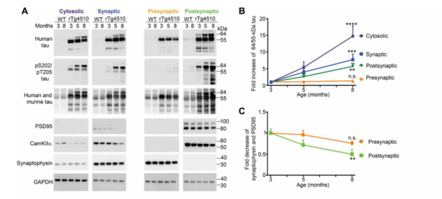
(Figure 1 A-C) Pathological tau protein accumulation in the postsynaptic compartment of AD patients
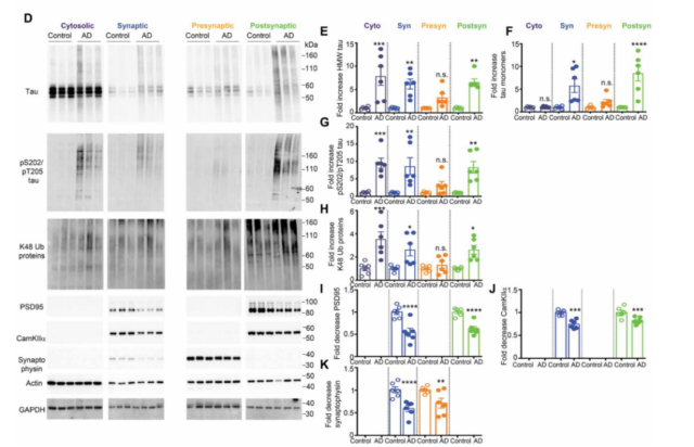
(Figure 1D-K) Pathological tau protein accumulation in the postsynaptic compartment of AD patients
Tau protein pathology has a basic feature, that is, tau protein with seed function will preferentially be transported through synapses and pass through adjacent brain regions.
Here, the author expressed the same amount of tau protein seeds extracted from mice at different disease stages in human embryonic kidney 293 DS1 cells, and found that compared with pre-synaptic tau seeds, post-synaptic tau seeds Tau seeds show high seed activity both in the early and late stages of the disease.
The same experiment was performed on the presynaptic and postsynaptic tau proteins isolated from the brain tissue of AD patients, and similar results were also observed. These results indicate that the tau in the postsynaptic compartment has more seed activity and more spreading potential.
Pituitary adenylate cyclase activating polypeptide (PACAP) type I receptor, referred to as PAC1R, is a Gs-G protein-coupled receptor mainly located on the postsynaptic membrane of neurons. It can activate the proteasome activity and tau protein degradation mediated by cAMP/PKA signaling pathway.
In the brain, after the ligand PACAP is released from the axon terminal, it acts by binding and activating the receptor PAC1R on the postsynaptic membrane. Here, the authors infected tau granules in primary neurons of tau mutant (P301S) mice, and observed that neuronal dendrites exhibited tau seed activity and insoluble tau accumulation, and PAC1R treatment can reduce dendrites The tau seed activity and insoluble tau can also significantly reduce the total post-synaptic tau protein content (Figure 2).

Figure 2 PACAP treatment can alleviate tau seed activity and insoluble tau accumulation in primary neurons in vitro (MC1 marks conformational tau protein, MAP2 marks neuronal dendrites)
Previous results indicate that enhancing the activity of PKA proteasome can promote the clearance of tau protein. The phosphorylated tau at serine 214 (pS204 tau) is a marker of activating cAMP/PKA signal transduction (in short, if the level of pS204 tau is high, the signaling pathway is activated).
Here, the authors found that pS214 tau protein was enriched in cytoplasm and presynaptic expression, while the PACAP treatment group significantly up-regulated the pS214 tau protein content in the cytoplasmic component of mouse neurons, suggesting PKA-dependent tau protein phosphorylation Occurs after PACAP treatment; the pS214 tau protein in the large presynaptic mice after PACAP treatment was significantly reduced, suggesting that PACAP treatment did not activate the presynaptic PKA.
The authors also found that PACAP treatment can significantly down-regulate mouse cytoplasm, pre-synaptic and post-synaptic proteasome-specific ubiquitination protein levels, and can also significantly down-regulate the level of post-synaptic receptor PAC1R. These results comprehensively suggest that PACA treatment can improve the tau protein pathology of mouse neuronal synapses.
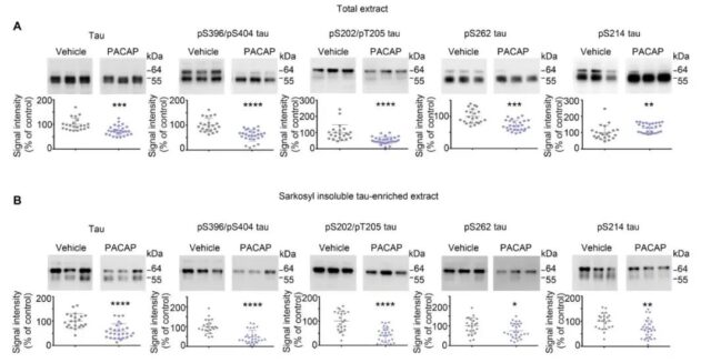
Figure 3 PACAP treatment can reduce tau protein pathology in rTg4510 mice
At the same time, the results also show that PACA treatment can significantly enhance the 26S proteasome activity and substrate degradation rate of the mouse synapse, and the phosphorylation level of the serine and threonine sites of the synaptic 26S proteasome also increases, and This phosphorylation is specific to PKA signaling (Figure 3).
These results suggest that PACA treatment can increase the activity of the postsynaptic proteasome by activating the PKA signaling pathway, and promote the hydrolysis of tau, thereby exerting a neuroprotective effect.
Indeed, further results show that PACA treatment can alleviate the tau protein lesions in the cortex and hippocampus of mice, which is reflected in the reduction of total tau protein, tau phosphorylated at multiple sites, and insoluble tau.
Finally, Morris water maze test and new object recognition test showed that PACAP treatment can significantly improve the spatial (learning) memory and declarative (situational) memory of the hippocampus of mice with early tau protein disease (rTg4510). These behavioral tests show that after receiving PACAP treatment, the tau protein pathology and memory ability of the mice have been improved.
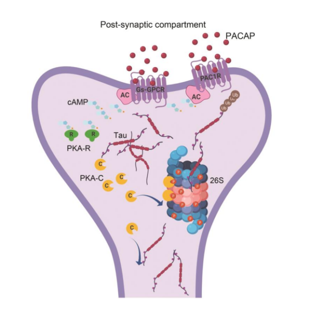
Figure 4 Work summary diagram: The mechanism of tau protein clearance in the postsynaptic compartment of neurons
The researchers demonstrated the biochemical properties of tau protein in the presynaptic and postsynaptic compartments of the brains of rTg4510 mice and AD patients after death by using subcellular separation methods (Figure 4). The postsynaptic compartment appears to contain pathological tau protein, which may make the neuronal synapse structure susceptible to the toxicity of tau protein.
The researchers used PACAP to target PAC1R to eliminate tau protein in the postsynaptic compartment of the mouse brain by increasing PKA signaling and enhancing proteasome activity (Figure 4). In PACAP-treated mice, the increase in proteasome activity in the postsynaptic compartment was associated with a pathological decrease in tau protein and an increase in cognitive ability (Figure 4).
In addition, this study also suggests that PKA phosphorylation of the proteasome is a better way to target the 26S proteasome enzyme activity. However, as an effector molecule of cAMP, PKA has a wide range of substrate proteins, which may be a long-term treatment for AD and other protein toxicity. Diseases that need attention.
Of course, there are still some unsolved problems in this study. For example, although PACAP treatment improves the cognitive ability of mice, PACAP has neurogenic, anti-apoptotic and neuroprotective properties, but its metabolic instability limits For its clinical application, it is necessary to develop PACAP derivatives or chemically modified small molecules to increase the stability of the enzyme while maintaining the selectivity and potency of PAC1R.
In conclusion, the research suggests that the method of targeted activation of PAC1R may be a potential strategy to prevent the accumulation of toxic tau protein and treat Alzheimer’s disease and other tau protein pathologies.
(source:internet, reference only)
Disclaimer of medicaltrend.org
Important Note: The information provided is for informational purposes only and should not be considered as medical advice.


