Revising Breast Screening Guidelines for Atypical Lesions
- Normal Liver Cells Found to Promote Cancer Metastasis to the Liver
- Nearly 80% Complete Remission: Breakthrough in ADC Anti-Tumor Treatment
- Vaccination Against Common Diseases May Prevent Dementia!
- New Alzheimer’s Disease (AD) Diagnosis and Staging Criteria
- Breakthrough in Alzheimer’s Disease: New Nasal Spray Halts Cognitive Decline by Targeting Toxic Protein
- Can the Tap Water at the Paris Olympics be Drunk Directly?
Revising Breast Screening Guidelines for Atypical Lesions
- Should China be held legally responsible for the US’s $18 trillion COVID losses?
- CT Radiation Exposure Linked to Blood Cancer in Children and Adolescents
- FDA has mandated a top-level black box warning for all marketed CAR-T therapies
- Can people with high blood pressure eat peanuts?
- What is the difference between dopamine and dobutamine?
- How long can the patient live after heart stent surgery?
Revising Breast Screening Guidelines for Atypical Lesions
Breast cancer is one of the most common malignant tumors globally. Screening can improve its early diagnosis, which is crucial for its treatment and prognosis.
However, besides detecting typical benign or malignant lesions, breast cancer screening can also identify some atypical lesions, including atypical ductal hyperplasia (ADH), flat epithelial atypia (FEA), atypical lobular hyperplasia (ALH), and lobular carcinoma in situ (LCIS).
These atypical lesions may not cause symptoms in a patient’s lifetime, potentially leading to overdiagnosis or overtreatment.
Atypical lesions refer to differences in cell morphology and tissue structure compared to the corresponding normal tissues, but this does not necessarily indicate a risk of malignancy. In the UK’s breast screening program, 5%-10% of screened individuals are diagnosed with atypical lesions. While these lesions are not malignant themselves, cancer cells can coexist with them. An analysis from a meta-analysis involving 13 studies and 1,759 female participants with a median follow-up of 15.7 years showed that the presence of atypical lesions increased the long-term risk of developing breast cancer by 4 times.
A recent prospective cohort study published in The BMJ analyzed big data on the number and types of breast cancer cases that occurred after detecting atypical lesions in every three-year breast screening in the UK.
The results showed that female participants who were detected with atypical lesions during breast cancer screening had similar grades, sizes, and lymph node involvement of subsequent invasive breast cancer compared to the entire screening population, with the same number of breast cancers on the same and opposite sides.
In summary, the vast majority of atypical lesions detected in breast screening may only represent risk factors rather than precursors to invasive breast cancer that require surgery in the short term.
Some guidelines currently recommend using vacuum-assisted core biopsy (a common minimally invasive surgical technique in breast surgery that uses a vacuum pump to remove masses) to remove atypical lesions, followed by annual breast molybdenum target monitoring. For atypical lesions with uncertain malignant potential (i.e., breast biopsies classified as B3), FEA and lobular tumors (including ALH and LCIS) are recommended to be removed by vacuum-assisted biopsy and then undergo 5 years of imaging surveillance; ADH is recommended to be removed by open surgery (monitoring can only replace surgical removal in special cases).
The current study analyzed data from a prospective cohort in the UK Sloane Atypia Project, which included 3,238 female participants diagnosed with atypical lesions after routine breast screening, covering information such as the type of atypical lesion, age at diagnosis, imaging features, biopsy methods, histological features, surgery, and adjuvant treatment.
Overall, the average age of the participants was 55.6 years. Among them, 1350 cases were ADH, 403 cases were FEA, 1101 cases were unspecified types of lobular neoplasia (LISN), and 384 cases were a mixture of ductal and lobular atypical lesions. Additionally, 2525 cases (78%) of patients diagnosed with atypical lesions had microcalcifications.
The analysis showed that between 2010 and 2015, the incidence of atypical lesions in women increased by 4 times, which was related to the introduction of digital mammography (DM) in breast screening centers from 2010 to 2013. The average follow-up time for these 3,238 atypical lesion participants was 5.9 years, of which 168 cases (5.2%) developed breast cancer (141 cases were invasive cancer; 27 cases were ductal carcinoma in situ [DCIS; stage 0 breast cancer, with a higher cure rate]).
The analysis indicated that the characteristics of invasive cancer in these participants were similar to those found in the general population through screening. The majority of invasive lesions were ≤20 mm in size and lymph node negative. Furthermore, the proportion of grade for these 141 invasive cancers was similar to those found in the general population through screening: 25 cases (17.7%) were grade 1; 69 cases (48.9%) were grade 2; 28 cases (19.9%) were grade 3; and 19 cases (13.5%) had no recorded grade.
After being diagnosed with atypical lesions, the cumulative incidence rates of invasive cancer in these participants were 0.95 cases per 1000 people in the first year and 14.2 cases per 1000 people in the third year (per 1000 people refers to every 1000 participants with atypical lesions). The paper emphasizes that the cumulative incidence rates of invasive cancer on the same side and opposite side in the third year after being diagnosed with atypical lesions were similar, at 7.7 cases per 1000 people and 6.5 cases per 1000 people, respectively. Although the data on the location of the lesions for invasive cancer on the same side were not complete, considering the number of invasive cancers on the opposite side, the researchers believe that many atypical lesions may not be precursors to subsequent breast cancer.
Moreover, the different surgical strategies (diagnostic surgical excision vs. secondary vacuum-assisted biopsy excision/vacuum-assisted core biopsy) for different types of atypical lesions did not affect the subsequent incidence of invasive cancer. In other words, vacuum-assisted core biopsy seems to be as safe as surgical excision for interventions targeting atypical lesions. It is worth mentioning that the analysis found that the risk of developing invasive cancer was similar for different types of atypical lesions, and there is currently no evidence to suggest that atypical lesions need to be stratified by subgroup for risk assessment.
The paper emphasizes that the short-term risk of developing breast cancer within 3 years after being diagnosed with atypical lesions is not high, and it may be overly diligent to have annual breast molybdenum target checks during this period. Instead, we may need to focus more on the long-term risk of developing breast cancer in atypical lesion participants 3-6 years later, when it may be necessary to have annual breast molybdenum target checks.
Overall, compared to the general population, atypical lesion participants have a lower risk of subsequent cancer incidence. In participants diagnosed with atypical lesions, there appears to be very few cases of cancer being missed in subsequent screenings. The paper emphasizes that atypical lesions may be risk factors for invasive cancer rather than precursors to invasive cancer. Therefore, it may not be necessary to have annual breast molybdenum target checks in the short term after being diagnosed with atypical lesions.
Revising Breast Screening Guidelines for Atypical Lesions
References:
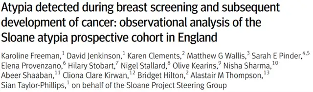
[1] Karoline Freeman et al. Atypia detected during breast screening and subsequent development of cancer: observational analysis of the Sloane atypia prospective cohort in England. The BMJ (2024). Doi: 10.1136/bmj-2023-077039
(source:internet, reference only)
Disclaimer of medicaltrend.org
Important Note: The information provided is for informational purposes only and should not be considered as medical advice.


