AJG special case: mixed cell liver cancer with clear boundaries
- Normal Liver Cells Found to Promote Cancer Metastasis to the Liver
- Nearly 80% Complete Remission: Breakthrough in ADC Anti-Tumor Treatment
- Vaccination Against Common Diseases May Prevent Dementia!
- New Alzheimer’s Disease (AD) Diagnosis and Staging Criteria
- Breakthrough in Alzheimer’s Disease: New Nasal Spray Halts Cognitive Decline by Targeting Toxic Protein
- Can the Tap Water at the Paris Olympics be Drunk Directly?
AJG special case: mixed cell liver cancer with clear boundaries
AJG special case: mixed cell liver cancer with clear boundaries. AJG special case: hepatocellular carcinoma and cholangiocarcinoma mixed cell liver cancer with clear boundaries.
Primary liver cancer can be divided into three categories: hepatocellular carcinoma (HCC), intrahepatic cholangiocarcinoma (ICC), and combined hepatocellular-cholangiocarcinoma (CHC).
Compared with the former two, CHC is relatively rare, accounting for about 0.4% to 14% of primary liver tumors. Because of its rarity and difficulty in preoperative diagnosis, research reports on this type of liver cancer are relatively scarce.
Recently, the American Journal of Gastroenterology (The American Journal of Gastroenterology, AJG, Proceedings of the American Society of Gastroenterology) published a special case online, describing a case that can be clearly distinguished in preoperative imaging, intraoperative gross, and postoperative pathology Of mixed cell liver cancer containing two components: hepatocellular carcinoma and intrahepatic cholangiocarcinoma.
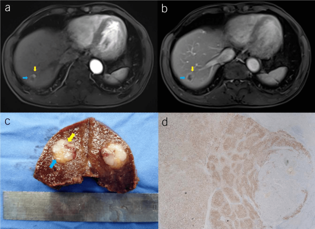
The study reported a 63-year-old male patient with a history of hepatitis B cirrhosis for 2 years. At the time of presentation, the AFP was 16.13ng/mL. The enhanced MRI of the liver showed that the liver segment VII showed a slightly longer oval T1 and long T2 signal, and the enhanced scan showed The ring continues to strengthen, and nodular enhancements can be seen in the ring. The lesions resemble a “ring”, and there is a clear boundary.
In most of the outer ring area (blue arrow), both the arterial phase and the venous phase are gradually strengthened, while in the area on the other side (yellow arrow), the typical fast-forward and fast manifestations can be seen, that is, the arterial phase and the venous phase. Eluted. No other abnormal findings were found on physical examination and laboratory examination.
Based on the size and location of the patient’s mass, the research team performed a fluorescence-navigated laparoscopic segment VII liver resection. The cross-section of the gross specimen after surgery shows that the lesion contains two visible tissues with clear boundaries. It is guessed that the larger part is hepatocellular carcinoma (yellow arrow), and the smaller part is intrahepatic cholangiocarcinoma (blue arrow). Colored arrow), and the postoperative pathology also confirmed this clinical judgment. It is also seen that the two components under the microscope are very significant differences and clear junctions.
Picture source: original document
CHC includes three subtypes:
- Type A: HCC and ICC exist separately in the liver;
- Type B: HCC and ICC are close but independent of each other;
- Type C: HCC and ICC are fused and interwoven. This patient should be type B.
There are few reports on magnetic resonance imaging studies of CHC, especially in this case, CHC that can be clearly distinguished in preoperative imaging, intraoperative gross, and postoperative pathology is very rare. Due to the particularity of histology and pathology, CHC has the characteristics of strong invasiveness and poor prognosis, but its clinical manifestations are similar to those of HCC and ICC and its vague imaging features. Preoperative diagnosis is difficult but important.
The preoperative imaging features of double-lesion liver cancer presented in this case deserve the attention and reference of clinical imaging experts.
Case summary
Hepatocellular-cholangiocyte mixed liver cancer is a rare but highly aggressive primary liver cancer, which is characterized by both hepatocellular carcinoma (HCC) and cholangiocarcinoma (CCA). The overall prognosis of the patient is poor, with a median overall survival of only 9 months reported in the literature.
Surgical resection is the preferred radical method. For unresectable cases, non-surgical treatment options include transarterial chemoembolization (TACE), stereotactic radiotherapy (SBRT), radiofrequency ablation, systemic chemotherapy, targeted therapy, and immunotherapy. However, there is still a lack of reliable efficacy data.
Small sample studies have shown that platinum-based chemotherapy (such as gemcitabine/cisplatin) is superior to tyrosine kinase inhibitors (such as sorafenib), but there is a lack of evidence from randomized controlled clinical studies. Due to the lack of treatment plans for specific diseases, and there is no standardized treatment plan, the diagnosis and treatment of patients with mixed liver cancer relies to a greater extent on precise and individualized methods, which guide follow-up treatment based on precise classification at the multi-omics level. At the same time, interdisciplinary full-process collaboration also plays a key role in the diagnosis and treatment of such patients.
(source:internet, reference only)
Disclaimer of medicaltrend.org


