Memory killer T cells may be activated in spleen During influenza infection
- Normal Liver Cells Found to Promote Cancer Metastasis to the Liver
- Nearly 80% Complete Remission: Breakthrough in ADC Anti-Tumor Treatment
- Vaccination Against Common Diseases May Prevent Dementia!
- New Alzheimer’s Disease (AD) Diagnosis and Staging Criteria
- Breakthrough in Alzheimer’s Disease: New Nasal Spray Halts Cognitive Decline by Targeting Toxic Protein
- Can the Tap Water at the Paris Olympics be Drunk Directly?
Memory killer T cells may be activated in spleen During influenza infection
- Should China be held legally responsible for the US’s $18 trillion COVID losses?
- CT Radiation Exposure Linked to Blood Cancer in Children and Adolescents
- FDA has mandated a top-level black box warning for all marketed CAR-T therapies
- Can people with high blood pressure eat peanuts?
- What is the difference between dopamine and dobutamine?
- How long can the patient live after heart stent surgery?
Memory killer T cells may be activated in the body’s spleen During influenza virus infection.
CD8+ T cells (killer T cells) are the “killer” of the immune system. Once activated, they will find and destroy cells infected by the virus or in a cancerous state.
And its activation often involves dendritic cells, which are the sentinels of the body’s immune system. For example, in a lung influenza virus infection, the migration of lung dendritic cells will capture a piece of viral antigen and then migrate from the lungs.
Go to the location of the original T cell and present the antigen to the CD8+ T cell, which will allow the T cell to know which cells need to be attacked.
 Image source: https://www.science.org/doi/10.1126/sciimmunol.abg6895
For a long time, scientists believed that the initiation site of influenza virus was limited to one anatomical site, that is, the lung draining mediastinal lymph nodes located between the lungs and the spine.
Recently, an article was published in the international journal Science Immunology entitled “Lung dendritic” In the research report on cells migrate to the spleen to prime long-lived TCF1hi memory CD8+ T cell precursors after influenza infection, scientists from the University of Alabama and other institutions challenged the fixed paradigm centered on lymph nodes through research.
Through research, we discovered an unnoticed extra part of CD8+ T cell function-the spleen.
This surprised the researchers, but it is very important because there is no lymphatic connection between the lung and the spleen; because the CD8+ T cells that are activated there are transcriptionally different, compared with the T cells in the lymph nodes , It may face another fate, and the CD8+ T cells in the lymph nodes may be ready to transform into effector T cells and return to the lung tissue to fight infection.
But those T cells that are stimulated in the spleen will produce precursors, and their ability to differentiate into long-lived stem-like memory T cells will be enhanced. Such memory cells can quickly respond to future influenza virus infections, thereby providing long-lasting The protective immunity.
Researcher Ballesteros-Tato said that now we have clarified that CD8+ T cells respond to the same antigen in different anatomical locations, so that cells with different functions can be produced, thus providing a new anatomical model to clarify infection How is the diversity of post T cells produced?
The results of this study show that the transport pathway of dendritic cells may connect the lungs and blood circulation. At the same time, the researchers also determined that the spleen is a major place where the precursors of long-lived memory T cells exist. The relevant research data is useful for the development of efficient vaccines.
Vaccination strategies and the development of therapeutic strategies to deal with respiratory diseases are both important. The researchers used mouse/influenza virus infection models to study and found that the activation effect in the spleen may be completed by the dendritic cells that migrated from the lymph nodes to the lungs. These cells enter the blood and return to the spleen. .
In addition to the transcriptional differences of T cells stimulated in the spleen, the “fate mapping experiment” also showed that CD8+ T cells stimulated in the spleen are longer-lived and can be compared with CD8+ T cells stimulated in integrated lymph nodes. Promote the establishment of a long-lived memory cell bank.
After further research, the researchers found that 45 days after infection, when the spleen stimulated CD8+ T cells and lymph nodes stimulated CD8+ T cells, there is no difference in phenotype, and the T cells stimulated in the spleen have another challenge to influenza virus. Stronger response ability, and can expand into a population of T effector cells that resist infection.
In the summary of experimental results that support this new paradigm, the researchers found that when dendritic cells cannot migrate from the lungs, or when lymph node drainage is inhibited, dendritic cells carrying lung-derived antigens cannot be found in the spleen. It accumulates in the spleen, thereby preventing the activation or excitation of T cells in the spleen.
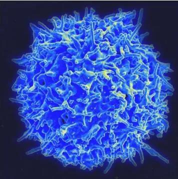 Scanning electron micrograph of T lymphocytes (also called T cells) in the immune system of a healthy donor.
Image source: NIAID
So how do migrating dendritic cells in the lungs get from the lymph nodes to the spleen? Researcher Ballesteros-Tato said that neither mediastinal lymph nodes nor lung tissue can be directly connected to the spleen through lymphatic ducts.
Therefore, the only possible way for migrating dendritic cells to enter the spleen from the mediastinal lymph nodes is through the thoracic duct.
This bile duct can lead the outflowing lymph fluid back into the blood circulation. At present, the knowledge about most stem-like CD8+ T cells comes from the study of tumor models or the study of chronic systemic viral infections; to date, there has not been any research to solve this problem, that is, these cells are in the respiratory virus.
How does the infection occur in the context of the infection.
In summary, the results of this article show that in addition to the lymph nodes draining from the lungs, the spleen may also be an excellent place for lung-derived dendritic cells to trigger a unique antiviral T cell response during influenza virus infection.
(source:internet, reference only)
Disclaimer of medicaltrend.org
Important Note: The information provided is for informational purposes only and should not be considered as medical advice.


