Moderna Team Detects No Uptake of mRNA-LNPs in Muscles at Injection Site
- Normal Liver Cells Found to Promote Cancer Metastasis to the Liver
- Nearly 80% Complete Remission: Breakthrough in ADC Anti-Tumor Treatment
- Vaccination Against Common Diseases May Prevent Dementia!
- New Alzheimer’s Disease (AD) Diagnosis and Staging Criteria
- Breakthrough in Alzheimer’s Disease: New Nasal Spray Halts Cognitive Decline by Targeting Toxic Protein
- Can the Tap Water at the Paris Olympics be Drunk Directly?
Moderna Team Detects No Uptake of mRNA-LNPs in Muscles at Injection Site
- Should China be held legally responsible for the US’s $18 trillion COVID losses?
- CT Radiation Exposure Linked to Blood Cancer in Children and Adolescents
- FDA has mandated a top-level black box warning for all marketed CAR-T therapies
- Can people with high blood pressure eat peanuts?
- What is the difference between dopamine and dobutamine?
- How long can the patient live after heart stent surgery?
Moderna Team Detects No Uptake of mRNA-LNPs in Muscles at Injection Site
Due to the presence of abundant RNases in bodily fluids, naked mRNA is highly unstable and degrades rapidly.
The inherent chemical properties, such as large size, hydrophilicity, and negative charge, make it difficult for mRNA to traverse cell membranes. Lipid Nanoparticles (LNPs) serve as an ideal delivery system, encapsulating mRNA to prevent degradation and facilitating its delivery from the injection site to target locations.
Once in the target tissue, LNPs are rapidly taken up by cells, allowing mRNA to escape into the cytoplasm and complete protein expression.
The human immune system consists of central and peripheral immune organs. Central immune organs, such as the thymus and bone marrow, are sites where immune cells are produced, differentiated, and matured.
The bone marrow is where B lymphocytes differentiate and mature, while the thymus is the site for T lymphocyte maturation.
Peripheral immune organs, known as secondary immune organs, are locations where mature T and B lymphocytes, along with other immune cells, settle and initiate immune responses.
These include lymph nodes (LNs), the spleen, and mucosal-associated tissues.
For mRNA vaccines to successfully trigger a robust immune response, it is crucial to ensure their smooth delivery to key immune organs. LNPs, with a size smaller than 200nm, interact with cells at the injection site and can flow back to lymph nodes through the lymphatic system.
Researchers believe that antigens encoded by mRNA vaccines reach draining lymph nodes through two pathways:
- Firstly, active transport by Antigen-Presenting Cells (APCs). mRNA-LNPs rapidly recruit immune cells at the injection site, triggering a natural immune response. APCs, such as dendritic cells (DCs) and macrophages, internalize mRNA-LNPs. Subsequently, APCs transport vaccine antigens produced inside the cells to draining lymph nodes (dLNs), initiating an adaptive immune response.
- Secondly, passive transport through the lymphatic system to draining lymph nodes. mRNA-LNPs flow back to draining lymph nodes through the lymphatic circulation and directly interact with APCs there.
In summary, the transient activation of the natural immune response by mRNA-LNPs ultimately leads to the activation of B lymphocytes and T lymphocytes in draining lymph nodes.
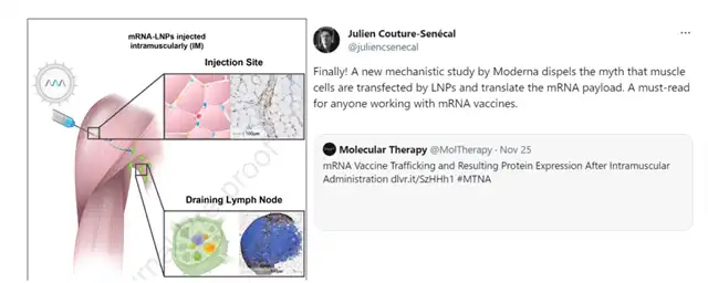
Intramuscular (IM) injection is the most common vaccination route. Recently, the Moderna team conducted a study on the delivery pathways of mRNA-LNPs vaccines after injection, quantitatively examining the dynamic changes of mRNA and encoded proteins in the injection site, lymph nodes, and specific tissues over time.
The related research is published in Molecular Therapy: Nucleic Acid: “mRNA Vaccine Trafficking and Resulting Protein Expression After Intramuscular Administration,” breaking previous perceptions by not observing mRNA or antigen protein expression in muscle fibers.
This important document is a must-read for mRNA vaccine practitioners, aiding in a better understanding of the delivery pathways of mRNA vaccines in the body.
Delivery pathway of mRNA-LNPs injected into the muscle of rodent animals
In mice, 3μg eGFP mRNA-LNPs were injected into the left quadriceps muscle. Protein expression and eGFP mRNA were detected using immunohistochemistry (IHC) and in situ hybridization (ISH) techniques. At the injection site, many neutrophils, macrophages, and a small number of lymphocytes infiltrated the connective and adipose tissues between muscles. At 6h/24h post-injection, mRNA and eGFP protein were observed in macrophages, fibroblasts, and adipocytes at the injection site. Surprisingly, eGFP mRNA or eGFP protein was not observed in muscle fibers at both time points.
Subsequently, researchers examined the levels of eGFP mRNA or eGFP protein in draining lymph nodes. eGFP protein was detected in the popliteal and inguinal lymph nodes. Inside the draining lymph nodes, eGFP protein was mainly expressed in macrophages in the subcapsular sinus and medullary sinus, reaching peak levels at 6h post-injection. At 24h post-injection, many eGFP mRNAs had disappeared in draining lymph nodes, with the remaining mRNA signals located in B-cell follicles and follicular interstitium. Additionally, a similar trend was observed in the levels of eGFP mRNA or eGFP protein in the injection site and draining lymph nodes of rats.
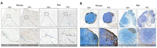
Detection of mRNA and protein expression in the injection site and draining lymph nodes of rodent animals (mice and rats) after muscle injection of mRNA-LNPs.
Delivery pathway of mRNA-LNPs injected into the muscle of NHPs
Considering the weight difference between rodents and non-human primates (NHPs), researchers increased the accumulation of eGFP mRNA-LNPs to 300μg in the NHP animal model. Tissues and plasma were collected at different times to detect eGFP mRNA concentrations. The distribution of the highest eGFP mRNA concentration in different immune tissues was as follows: spleen > draining lymph nodes > injection site > plasma > liver. After 24h post-injection, eGFP mRNA was completely undetectable in the injection site, lymph nodes, and liver, with a significant decrease in mRNA in plasma and spleen. These data indicate that free mRNA in immune tissues is quickly degraded.
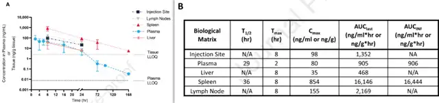
Dynamic changes in mRNA concentration in the injection site and various immune tissues over time after muscle injection in NHPs.
In the NHP injection site, eGFP mRNA signals peaked at 8h post-injection. Over time, eGFP mRNA signals decreased, approaching near disappearance at 72h post-injection. Consistent with rodents, eGFP mRNA signals at the NHP injection site were mainly distributed in inflamed macrophages, fibroblasts, and adipocytes. No eGFP mRNA signals were detected in muscle fibers. Similarly, cells expressing eGFP protein were mainly macrophages, fibroblasts, and adipocytes. Additionally, eGFP protein expression was observed in endothelial cells at the NHP injection site, a situation not observed in rodent injection sites.
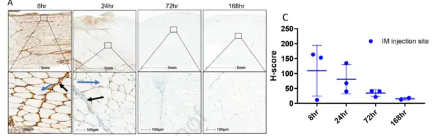
Dynamic changes in eGFP mRNA levels in the NHP injection site over time.
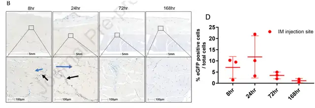
Dynamic changes in eGFP protein levels in the NHP injection site over time.
The eGFP mRNA signal in the iliac lymph nodes of NHPs was higher than in popliteal and inguinal lymph nodes. At 8h post-injection, eGFP mRNA signals in the subcapsular sinus and medullary sinus of draining lymph nodes were predominant. Subsequently, a small amount of eGFP mRNA was detected in B-cell follicles.
In the medulla, eGFP protein signals were observed in tissue cells, reticular cells, and dendritic cells. Additionally, eGFP protein signals were observed in the sinus endothelial cells in the sinus and subcapsular sinus. In the marginal zone of follicles, few tissue cells and reticular cells also expressed eGFP protein.
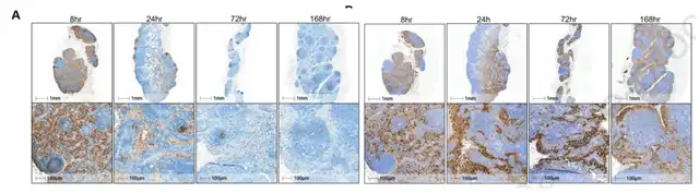
Dynamic changes in eGFP mRNA (A) and eGFP protein (B) levels in NHP draining lymph nodes over time.
Conclusion
After mRNA-LNPs were injected into the muscle, researchers demonstrated that in both NHPs and rodents, mRNA distribution and encoded protein expression mainly occurred in infiltrated immune cells at the injection site, followed by appearance in draining lymph nodes.
High levels of protein expression were observed in macrophages in the subcapsular and medullary sinuses inside draining lymph nodes. Interestingly, whether in NHPs or rodents, mRNA and protein expression were not detected in muscle fibers at the injection site, indicating that mRNA-LNP did not transfect muscle cells.
These data will help optimize the LNP delivery system, providing crucial guidance for the development of mRNA vaccines.
Moderna Team Detects No Uptake of mRNA-LNPs in Muscles at Injection Site
(source:internetGcHy1lnM_Cff6Y_1f6PASw, reference only)
Disclaimer of medicaltrend.org
Important Note: The information provided is for informational purposes only and should not be considered as medical advice.


