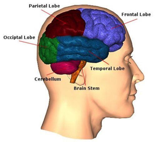
Classification and damage of delayed intracerebral hematoma
- Normal Liver Cells Found to Promote Cancer Metastasis to the Liver
- Nearly 80% Complete Remission: Breakthrough in ADC Anti-Tumor Treatment
- Vaccination Against Common Diseases May Prevent Dementia!
- New Alzheimer’s Disease (AD) Diagnosis and Staging Criteria
- Breakthrough in Alzheimer’s Disease: New Nasal Spray Halts Cognitive Decline by Targeting Toxic Protein
- Can the Tap Water at the Paris Olympics be Drunk Directly?
Classification and damage of delayed intracerebral hematoma
Classification and damage of delayed intracerebral hematoma. Systemic coagulation dysfunction may also be related to the formation of delayed traumatic brain hematoma.
Intracerebral hematomas are divided into three categories:
- Acute hematoma, intracerebral hematoma that occurs immediately after injury
- Delayed hematoma, intracerebral hematoma that appears several days to several weeks after injury
- Chronic hematoma, intracerebral hematoma that occurs several months after injury
The incidence of delayed traumatic intracerebral hematoma accounts for 0.3% to 3.5% of all patients with traumatic brain injury. Clinical statistics also show that the incidence of delayed traumatic intracerebral hematoma in patients with severe head injury has increased significantly, accounting for about 6.5% -8.7% of patients with severe head injury.
Cerebral ischemic damage, liquefaction and necrosis of brain tissue, and cerebral vascular rupture and hemorrhage caused by secondary vasospasm after craniocerebral trauma are the main causes of delayed traumatic intracerebral hematoma.
Delayed traumatic intracerebral hematoma usually occurs in the area of the primary brain contusion, suggesting that the primary brain contusion is related to the mechanism of delayed traumatic intracerebral hematoma. Primary brain injury will lead to local cerebrovascular self-regulation dysfunction, and then local cerebral congestion and hemorrhage will gradually form delayed intracerebral hematoma.
However, some people found through CT scan and autopsy that some delayed traumatic intracerebral hematomas are related to cerebral ischemic infarction, and there is usually no obvious primary brain contusion in the deep parenchyma of the brain. Diffuse axonal injury in deep brain parenchyma may also be related to the pathogenesis of delayed traumatic intracerebral hematoma.
Delayed traumatic intracerebral hematomas are also common after removal of intracranial and extracerebral hematomas or after ventricle puncture and cerebrospinal fluid drainage. As the intracranial pressure drops sharply after hematoma removal, the brain tissue is displaced, causing the thrombus formed at the damage of the cerebral parenchymal blood vessels to fall off, causing the blood vessels to rebleed and form intracerebral hematomas. In addition, due to the sharp decrease in intracranial pressure, the blood vessels in the primary brain contusion and laceration area were pulled torn and bleeds due to displacement, forming intracerebral hematomas.
Others believe that the blood vessels in the primary brain contusion and laceration area are gradually damaged and ruptured due to secondary ischemia and hypoxia for a long time, and then delayed diffuse hemorrhage occurs, forming intracerebral hematoma. When the intracranial pressure drops sharply, the chance and speed of bleeding increase significantly, and delayed traumatic intracerebral hematoma is more likely to occur.
It is common clinically after removal of intracranial hematoma, after ventricular shunt or after ventricular drainage. In addition, systemic coagulopathy may also be related to the formation of delayed traumatic brain hematoma.
(source:internet, reference only)
Disclaimer of medicaltrend.org


