Breast cancer early screening: 3D tomographic mammography machine
- Normal Liver Cells Found to Promote Cancer Metastasis to the Liver
- Nearly 80% Complete Remission: Breakthrough in ADC Anti-Tumor Treatment
- Vaccination Against Common Diseases May Prevent Dementia!
- New Alzheimer’s Disease (AD) Diagnosis and Staging Criteria
- Breakthrough in Alzheimer’s Disease: New Nasal Spray Halts Cognitive Decline by Targeting Toxic Protein
- Can the Tap Water at the Paris Olympics be Drunk Directly?
Breast cancer early screening: 3D tomographic mammography machine.
Breast cancer early screening: 3D tomographic mammography machine. How is mammography performed?
A device for early detection and diagnosis of breast diseases: a digital three-dimensional (3D) tomographic mammography machine.
Mammography is currently the first choice and the simplest and most reliable non-invasive detection method for the diagnosis of breast diseases, and it has the characteristics of comprehensive, intuitive, simple operation, safety and reasonable cost.
Compared with the traditional digital breast machine, the “new skills” of this device
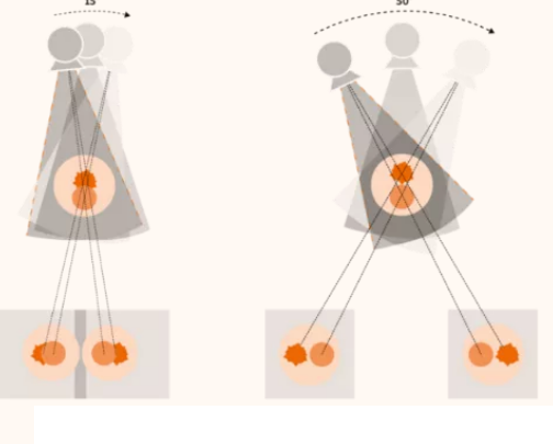
Compared with the traditional two-dimensional (2D) photography, the three-dimensional (3D) tomography system can increase the tumor detection rate by 25%, increase the detection rate of invasive cancer by 40%, increase the positive predictive value by 50%, and reduce by 20%. % Recall rate. Dense glands have clear layers, reducing false positive rates.
The device also has a three-dimensional stereotactic biopsy system, which can easily and accurately perform preoperative guide wire positioning and minimally invasive breast biopsy. Using this method can ensure accurate positioning of the operation, help preoperative qualitative tumors, and minimize the operation Scope, reduce trauma.
How is mammography performed?
The standard inspection procedure includes X-ray projections of two breasts in two positions, a total of four exposures, and breast compression imaging.
The patient is standing in front of the mammography machine. The technician places the breast on the pallet, and then slowly compresses the compression plate, flattening the breast slightly and then exposing it. Compression forces the patient to feel a certain amount of pressure, sometimes mild pain, but each time of compression is generally less than one minute.
The high-permeability honeycomb grid technology improves the image contrast by reducing the influence of scattered rays, and can reduce the radiation dose by more than 30%.
Why does mammography need to be squeezed?
Squeezing brings the most important benefits to the subject:
First, the radiation dose can be greatly reduced.
Second, improve image clarity and ensure the quality of diagnosis. The discomfort (including pain) caused by breast squeezing is within a tolerable range. The actual maximum squeezing state of each film only lasts for a few seconds, generally less than 5 seconds.
Who is recommended for mammography?
Young women aged 25-35 are clinically suspected of malignant breast lesions and should undergo mammography.
Women 35-40 years old and over 55 years old have a mammogram every 1.5 to 2 years. It is recommended that the inspection report be kept as basic information, so that it can be compared with the results of future mammography examinations, which will help to find the changes.
Women aged 40-55 are recommended to have a mammogram once a year.
Regardless of the age of women, if they find breast tumors for the first time, they should go to a regular hospital breast specialist to rule out the possibility of breast cancer.
Matters needing attention during mammography examination:
1. Take off the shirt, expose the upper body, fully expose both breasts, and take pictures of the breasts.
2. Before the examination, the doctor should be informed of the medical history. Pregnant and breastfeeding women are not recommended to undergo mammography.
3. Try to avoid 3-5 days before and after the menstrual period, and the best check-up time is 1-2 weeks after the menstrual period.
4. Breast tissue will bear greater pressure during slow pressure. Please keep your body calm during the filming until the pressure is relieved.
(source:internet, reference only)
Disclaimer of medicaltrend.org


