The types and related characteristics of various anemias
- Normal Liver Cells Found to Promote Cancer Metastasis to the Liver
- Nearly 80% Complete Remission: Breakthrough in ADC Anti-Tumor Treatment
- Vaccination Against Common Diseases May Prevent Dementia!
- New Alzheimer’s Disease (AD) Diagnosis and Staging Criteria
- Breakthrough in Alzheimer’s Disease: New Nasal Spray Halts Cognitive Decline by Targeting Toxic Protein
- Can the Tap Water at the Paris Olympics be Drunk Directly?
The types and related characteristics of various anemias
The types and related characteristics of various anemias. Iron deficiency anemia, aplastic anemia, hemolytic anemia, megaloblastic anemia。
Megaloblastic anemia: A group of anemias caused by deoxyribonucleic acid (DNA) synthesis disorders. It is mainly caused by the lack of vitamin B12 or folic acid in the body. It can also be caused by inherited or acquired DNA synthesis disorders such as drugs.
Anemia?
Anemia: The red blood cell count, hemoglobin amount and hematocrit in a certain volume of circulating blood are all lower than normal standards. Among them, hemoglobin is the most important, adult males are less than 120g/L (12.0g/dl), and adult women are less than 110g/L (11.0/dl), which is generally considered anemia.
Common types of anemia:
Iron deficiency anemia
Iron deficiency anemia (IDA) refers to anemia that occurs due to the exhaustion of iron storage in the body and the inability to meet the needs of normal red blood cell production. Before the production of red blood cells is restricted, the iron storage in the body has been exhausted, and this is called iron deficiency.
Iron-deficiency anemia is the most common type of anemia, which mostly occurs in young children, pregnant and lactating women, chronic blood loss and gastrointestinal absorption dysfunction.
Blood picture: hemoglobin and red blood cells are reduced, and the hemoglobin reduction is more obvious. There is no obvious abnormality in the morphology of red blood cells in mild anemia, and the volume of red blood cells is reduced when anemia is above moderate, and the central lightly stained area is enlarged. In severe cases, the red blood cells can be ring-shaped, and there are pleochroic red blood cells and stippling red blood cells. The reticulocytes are slightly increased or normal. The white blood cell count and classification are generally normal. The platelet count is generally normal.
Bone marrow picture: The hyperplasia is obviously active. The ratio of grain to red is reduced. The red blood cell line is obviously proliferating, mainly in the middle and late young red blood cells. Immature red blood cells are small in size, have irregular edges, small and dense nuclei, and a small amount of cytoplasm. They are slightly alkaline due to insufficient hemoglobin synthesis. Mature red blood cells are small in size, and the lightly stained area in the center is enlarged. In severe cases, they can be ring-shaped red blood cells. Easily see pleochroic red blood cells. The granulocytic cell line is relatively reduced, but the proportion and cell morphology of each stage are roughly normal. The megakaryocyte cell line is normal.
Aplastic anemia
Aplastic anemia (AA) is a kind of physical, chemical, biological or unknown factors that cause severe damage to bone marrow hematopoietic stem cells and bone marrow microenvironment, resulting in reduced or failure of bone marrow hematopoietic function. Pancytopenia is one of the main manifestations. Group syndrome.
Acute aplastic anemia:
Blood picture: Red blood cells and hemoglobin are mostly severely reduced, and the two decrease in parallel, which is normal cell orthochromatic anemia. The white blood cells are severely reduced, and the neutrophil jealousy is reduced while the lymphocytes are relatively increased during classification. Platelets are severely reduced, and reticulocytes are severely reduced.
Bone marrow picture: Because the red bone marrow pathology of aplastic anemia is extensive and replaced by adipose tissue, it is not easy to obtain bone marrow components during puncture, and the fat droplets on the smear are obviously increased. Therefore, it often requires multiple and multi-site punctures to be diagnosed.
The main characteristics are: bone marrow hyperplasia is mostly severely reduced. Both the granulocyte and red lines were severely reduced. The granulocyte line was mainly mature granulocytes, and the red blood cell line was mainly medium and late young red. The staining of cell morphology was roughly normal. The lymphocytes are relatively increased. Plasma cells, reticulocytes, basophils and other non-hematopoietic cells increase, and those that appear in piles are “non-hematopoietic cell clusters.” Megakaryocytes are significantly reduced, and most of them do not see megakaryocytes except occasionally in some cases. There was no obvious change in the morphology and staining of mature red blood cells.
Chronic aplastic anemia:
Blood picture: Red blood cells, hemoglobin, white blood cells and platelets are moderately or severely reduced, mostly normal cells, orthochromic anemia. Leukopenia, the degree of neutropenia is not as good as the acute type, and the lymphocytes are relatively increased. Thrombocytopenia, and abnormal morphology can be seen, such as small size, irregular shape, and decreased intracytoplasmic granules. The absolute value of reticulocytes is still lower than normal.
Bone marrow picture: Chronic aplastic anemia is due to concentric damage to the bone marrow and modern compensatory hematopoietic foci. Therefore, the puncture findings are not consistent, and multiple and multi-site punctures are needed for diagnosis.
Main features: Bone marrow is mostly dysplasia, and when compensatory hematopoietic foci occurs, it can show that the hyperplasia is active or even obviously active. The total percentage of granulocytes is normal or decreased. Lymphocytes are relatively increased, but non-hematopoietic cells are less than acute aplastic anemia. Megakaryocytes are significantly reduced, even when bone marrow hyperplasia is good, megakaryocytes are also reduced, so this point is one of the very important conditions for the diagnosis of aplastic anemia. Morphological staining of mature red blood cells was roughly normal.
Hemolytic anemia
Hemolytic anemia: refers to a type of anemia that occurs when the destruction of red blood cells is accelerated and the hematopoietic function of the bone marrow is insufficient. If the bone marrow can increase red blood cell production, enough to compensate for the shortened survival of red blood cells, anemia will not occur. This state is called compensatory hemolytic disease
Blood picture: Red blood cells and hemoglobin are decreased, and the two are mostly decreased in parallel, and MCV can be seen to increase. The white blood cell count often increases, and the nucleus shifts to the left. Platelets may show increased reactivity. The size of mature red blood cells is uneven, large red blood cells are easy to see, pleochrotropic red blood cells increase, and immature red blood cells appear. The reticulocytes increased significantly.
Bone marrow appearance: Bone marrow hyperplasia is obviously active. The red blood cell line proliferates significantly, the primitive and early young red blood cells increase, but the middle and late young red blood cells are still the main increase, and the cell morphology staining is not abnormal. The total percentage of granulocytes can be relatively reduced, but the proportion of each stage and the staining of cell morphology are generally normal. The ratio of grain to red is significantly reduced or even inverted. Megakaryocytes are normal or increased. The morphological changes of mature red blood cells were the same as those of peripheral blood.
Megaloblastic anemia
Megaloblastic anemia: A group of anemias caused by deoxyribonucleic acid (DNA) synthesis disorders. It is mainly caused by the lack of vitamin B12 or folic acid in the body. It can also be caused by inherited or acquired DNA synthesis disorders such as drugs.
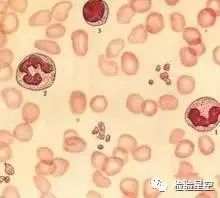
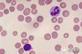
Blood picture: It is large cell orthochromic anemia (MCV>100fl), and the blood picture often shows pancytopenia. Neutrophils and platelets can be reduced, but it is less severe than anemia. Most of the large oval red blood cells can be seen in the blood smear, and the neutrophils have too many lobes, and there may be five or more lobes. Occasionally, huge platelets can be seen. The reticulocyte count is normal or slightly increased.
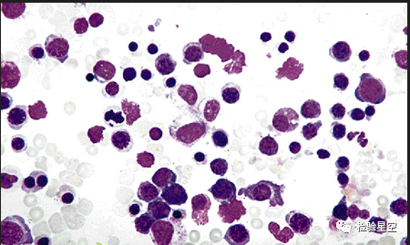
Bone marrow phenomenon: the bone marrow is proliferating actively, and the red blood cell line proliferation is significantly increased. All cell lines have megaloblasts, and the red blood cell line is the most significant. The cells of each stage of the erythroid are larger than normal, the cytoplasm is more mature than the nucleus, and the nuclear chromatin is scattered and concentrated. Similar morphological changes can be seen in granulocytes and megakaryocyte cell lines, especially in late immature and rod-shaped granulocytes. The morphological changes of mature red blood cells were the same as those of peripheral blood.

(source:internet, reference only)
Disclaimer of medicaltrend.org


